
1. Why lipoprotein(a) matters
Imagine carrying a cardiovascular risk factor that is largely set before birth, changes little through adult life, is not included in most standard lipid panels, and may remain undiscovered until coronary plaque or a cardiovascular event appears. That risk factor is lipoprotein(a).
Lp(a) was described by Kåre Berg in 1963. For decades it remained a specialist laboratory finding because measurement was inconsistent, the biology was complex, and there was no specific treatment. The scientific position is now very different. Large prospective cohorts show a continuous association between higher Lp(a) and myocardial infarction, ischemic stroke, peripheral artery disease and aortic stenosis. Genetic variants that raise Lp(a) also raise disease risk, strengthening the case that Lp(a) is causal rather than merely correlated with disease. [1,6-12]
The clinical relevance of Lp(a) is often easiest to understand as residual risk. A person may have an apparently satisfactory LDL-C result yet still carry substantial inherited risk from Lp(a). Conversely, an elevated Lp(a) concentration does not mean that an event is inevitable. Absolute risk depends on the cumulative burden of all causal exposures over time, including the total number of ApoB-containing particles, smoking, blood pressure, glucose metabolism and whether plaque is already present.
This distinction matters because the goal is not to create anxiety around an unchangeable gene. The goal is to detect a risk amplifier early enough to intensify the factors that are modifiable. The higher the Lp(a), the stronger the rationale to avoid years of unnecessary exposure to elevated LDL-C or ApoB, untreated hypertension, smoking or diabetes.
Lp(a) is best viewed as a risk amplifier, not as a stand-alone diagnosis. The same Lp(a) concentration has different consequences in a healthy 30-year-old with low ApoB and no smoking than in a 65-year-old with diabetes, CAC 400 and established vascular disease.
2. Structure, genetics and measurement
2.1 What exactly is an Lp(a) particle?
An Lp(a) particle contains an LDL-like lipid core and one molecule of ApoB-100, the same structural protein found on LDL and other atherogenic lipoproteins. Its defining feature is apolipoprotein(a), or apo(a), which is attached to ApoB-100 by a disulfide bond. Apo(a) is structurally related to plasminogen and contains repeated looped domains known as kringles. [2-6]
The number of kringle IV type 2 repeats varies widely. This copy-number variation produces apo(a) proteins of different sizes. In general, smaller apo(a) isoforms are secreted more efficiently and are associated with higher circulating Lp(a), although additional LPA variants also contribute. This molecular heterogeneity is one reason measurement is more difficult than measuring LDL-C.
Lp(a) is enriched in oxidized phospholipids. These biologically active lipids appear to contribute to endothelial activation, monocyte recruitment, arterial inflammation and calcific signaling in the aortic valve. The particle therefore combines the cholesterol-carrying properties of an ApoB particle with additional inflammatory and potentially thrombogenic biology. [9-13]
2.2 Why lifestyle changes Lp(a) very little
Most between-person variation in Lp(a) is explained by the LPA gene. Weight loss, a Mediterranean-style diet, exercise, improved sleep and smoking cessation are fundamental for cardiovascular health, but they generally do not lower Lp(a) to a clinically important degree. This is not a reason to dismiss lifestyle. It means lifestyle reduces the other components of risk rather than rewriting the inherited Lp(a) concentration.
Lp(a) can vary modestly with acute inflammation, significant kidney or liver disease, pregnancy, menopause and major hormonal change. A result obtained during an unstable medical period may occasionally deserve confirmation after recovery. For most adults, however, one well-performed lifetime measurement is sufficient to identify inherited exposure. [3-5]
2.3 mg/dL and nmol/L are not interchangeable
Mass assays report mg/dL and measure the mass of the complete particle. Molar assays report nmol/L and estimate particle concentration. Because apo(a) isoform size differs between people, particles do not all have the same mass. A fixed multiplier therefore cannot accurately convert every individual result from mg/dL to nmol/L or in the opposite direction.
Clinical interpretation should use the unit reported by the laboratory and the threshold used by the relevant guideline or study. When available, an isoform-insensitive assay calibrated to recognized reference material and reported in nmol/L is preferred. Historical studies and many clinical laboratories still use mg/dL, so both units will remain common. [3-5]
| Interpretive zone | mg/dL | nmol/L | How to use it |
|---|---|---|---|
| Lower / rule-out zone | <30 | <75 | Major Lp(a)-related excess risk is less likely, but total risk still depends on all other factors. |
| Intermediate gray zone | 30-49 | 75-124 | Interpret with age, family history, ApoB, diabetes, smoking, blood pressure and imaging. |
| Risk-enhancing zone | ≥50 | Commonly ≥125; ESC/EAS 2025 uses >105 | Supports more intensive global risk assessment and lower tolerance for other uncontrolled risk factors. |
| Very high concentration | ≈180 or higher | Assay dependent | May confer lifetime risk comparable to heterozygous familial hypercholesterolemia in some populations. |
Myth: 50 mg/dL always equals 125 nmol/L. Fact: those paired thresholds are pragmatic guideline anchors, not a valid person-specific conversion. Apo(a) size and assay design prevent a universal conversion formula.
2.4 Who should be tested?
The direction of modern guidance is clear: measure Lp(a) at least once in adulthood. Testing is especially important in premature ASCVD, familial hypercholesterolemia, recurrent events despite apparently good LDL-C control, calcific aortic stenosis, unexplained premature ischemic stroke, and families with early coronary disease or known high Lp(a). [3-5,23-24]
Because Lp(a) is strongly inherited, a markedly elevated result has implications for first-degree relatives. Cascade testing can identify children, siblings and parents who may benefit from earlier attention to LDL-C, smoking, blood pressure and other modifiable risks. Testing a child is particularly reasonable when there is familial hypercholesterolemia, very premature cardiovascular disease in the family, or a parent with markedly elevated Lp(a); interpretation should be coordinated with a clinician experienced in pediatric lipids.
3. How elevated Lp(a) causes disease
3.1 Atherogenic: it is an ApoB-containing particle
Atherosclerosis begins when ApoB-containing particles enter and are retained in the arterial wall. Each Lp(a) particle carries one ApoB molecule and can contribute cholesterol to plaque. Genetic analyses suggest that, per particle, Lp(a) may be substantially more atherogenic than ordinary LDL, although LDL particles are usually far more abundant and therefore remain the dominant modifiable driver in most people. [9,25-26]
This distinction explains why ApoB and Lp(a) are complementary. ApoB counts all circulating atherogenic particles, including each Lp(a) particle, while an Lp(a) assay identifies the inherited subset with apo(a)-related biology. A person can have a favorable ApoB result and still have a clinically important Lp(a), but lowering ApoB reduces the much larger background population of atherogenic particles with which Lp(a) interacts.
3.2 Pro-inflammatory: oxidized phospholipids
Lp(a) is a major carrier of oxidized phospholipids. Human mechanistic studies show that these lipids can promote inflammatory monocyte responses and arterial-wall inflammation. They may facilitate endothelial dysfunction, immune-cell recruitment and evolution of lipid-rich plaque. [10]
It is important not to equate this local vascular biology with a high blood hs-CRP in every patient. hs-CRP is a systemic marker, whereas Lp(a)-related oxidized phospholipids can act within plaque even when hs-CRP is low. A low hs-CRP is favorable but does not neutralize a high Lp(a).
3.3 Pro-thrombotic biology: plausible, but often overstated
Apo(a) resembles plasminogen, the precursor of the fibrinolytic enzyme plasmin. This structural similarity has generated a plausible hypothesis that Lp(a) may impair fibrinolysis or promote thrombosis. Experimental data support several prothrombotic pathways, but clinical evidence is stronger for arterial atherothrombosis than for routine venous thrombosis. It is therefore reasonable to describe Lp(a) as potentially prothrombotic, but not to assume that every person with high Lp(a) requires antiplatelet therapy.
3.4 Pro-calcific: the aortic valve
The causal link between LPA variation and calcific aortic valve disease is among the strongest findings in Lp(a) research. Lp(a) and its oxidized phospholipid cargo can stimulate osteogenic pathways in valvular tissue. Higher Lp(a) is associated with aortic-valve calcification, faster hemodynamic progression in some cohorts, and increased risk of clinical aortic stenosis. [11-13]
No lipid-lowering drug has yet been proven to stop established calcific aortic stenosis. The emerging Lp(a)-lowering era may eventually provide a mechanism-specific prevention strategy, but this remains an outcomes question rather than a current indication.
4. Interpreting cardiovascular risk
4.1 Risk is continuous, not a cliff at 50 mg/dL
Guidelines use practical thresholds, but biology does not switch on at a single number. Population risk rises progressively across the Lp(a) distribution. The absolute effect is greatest when baseline risk is already high. Thus, a very elevated Lp(a) has more immediate implications in someone with established plaque, diabetes or smoking than in a young person with otherwise optimal health.
Relative risk and absolute risk should not be confused. A biomarker can increase relative risk while the short-term absolute event probability remains low. This is particularly relevant in younger adults and in people with CAC zero. Lifetime exposure still matters, but the urgency and intensity of therapy should be proportional to overall risk.
4.2 Lp(a), LDL-C and ApoB
LDL-C estimates the cholesterol mass carried in LDL particles; ApoB estimates the total number of atherogenic particles; Lp(a) measures a genetically distinct subset. When LDL-C and ApoB are discordant, ApoB often provides a clearer representation of particle burden. When Lp(a) is high, measured LDL-C also includes some cholesterol carried inside Lp(a), which can complicate interpretation at very high concentrations.
The practical response is not to subtract uncertain amounts of Lp(a)-cholesterol and ignore LDL-C. It is to use LDL-C, non-HDL-C and ApoB together, recognize that Lp(a) adds risk, and lower the modifiable ApoB burden to a level appropriate for the patient's risk category. [25-26]
Causal ApoB exposure and causal Lp(a) exposure are additive, not mutually exclusive. Lowering LDL-C and ApoB remains beneficial even when the major inherited abnormality is Lp(a).
4.3 Family history and age
Family history can reveal cumulative genetic risk that a 10-year calculator underestimates. High Lp(a), familial hypercholesterolemia and polygenic susceptibility can cluster in the same family. A parent or sibling with myocardial infarction at an unusually young age should lower the threshold for Lp(a) testing and for early control of LDL-C.
Age has two meanings. It raises short-term risk because plaque burden generally accumulates with time, but it also determines how much future exposure can still be prevented. Identifying high Lp(a) at age 25 may be more useful than discovering it after an event at age 65, even if the young adult's 10-year risk is low.
5. Coronary artery calcium: powerful but incomplete
5.1 What CAC measures
A coronary calcium scan is a noncontrast CT examination that detects macroscopic calcification within coronary atherosclerotic plaque. The Agatston score combines the area and density of calcium. CAC is not calcium from food, vitamin D or a high blood calcium level. It is a marker of the coronary artery's accumulated response to atherosclerotic injury.
CAC is powerful because calcified coronary plaque is highly specific for atherosclerosis and because total calcium burden strongly predicts events. The test is quick, uses no iodinated contrast, and generally involves a low radiation dose. It is most useful when the result can change a prevention decision, especially in asymptomatic adults whose statin decision remains uncertain after clinical risk assessment. [15,23]
5.2 What CAC does not measure
CAC does not directly show the lumen, stenosis or noncalcified plaque. Early or lipid-rich plaque may exist before sufficient calcium is present to register on a calcium scan. CAC zero therefore means no detectable calcified plaque, not necessarily no atherosclerosis. The distinction is especially important in younger people, symptomatic patients and selected individuals with strong risk enhancers.
At the same time, the limitations of CAC should not be used to dismiss a zero score. In asymptomatic primary-prevention populations, CAC zero is one of the strongest available markers of low near-term coronary risk. The correct interpretation is 'reassuring but not invulnerable,' not 'perfectly healthy arteries' and not 'the scan missed everything.'
| CAC score | Typical interpretation | Practical implication |
|---|---|---|
| 0 | No detectable calcified plaque | Strongly reassuring for near-term risk in asymptomatic adults; exceptions include smoking, diabetes, strong family history, severe hypercholesterolemia or symptoms. |
| 1-99 | Definite but generally mild calcified atherosclerosis | Supports active risk-factor treatment; age and percentile matter. |
| 100-299 | Moderate plaque burden | Usually supports intensive prevention and lower LDL-C/ApoB targets. |
| ≥300 | High plaque burden | Risk can approach secondary-prevention levels in some cohorts; management is typically aggressive. |
| ≥1000 | Very extensive calcified atherosclerosis | Very high event risk; evaluate for symptoms and optimize all preventive therapy. |
5.3 How CAC and Lp(a) work together
Lp(a) and CAC are complementary. Lp(a) is a lifelong inherited exposure; CAC is evidence that coronary atherosclerosis has developed. In a combined analysis of MESA and the Dallas Heart Study, elevated Lp(a) and CAC were independently associated with ASCVD. Participants with elevated Lp(a) and CAC at least 100 had the highest risk, with an adjusted hazard ratio of 4.71 compared with those who had neither marker. [15]
The same study found that elevated Lp(a) with CAC zero was not associated with a statistically significant increase in events compared with non-elevated Lp(a) and CAC zero. This does not prove that Lp(a) becomes harmless. It shows that CAC zero can dominate near-term risk stratification in asymptomatic adults. Lp(a) remains relevant to lifetime prevention, family testing and the decision not to tolerate high LDL-C for decades.
| Lp(a) | CAC | Interpretive message |
|---|---|---|
| Not elevated | 0 | Low near-term coronary risk; maintain healthy prevention and reassess only if clinically useful. |
| Elevated | 0 | Near-term risk is often still low, but inherited lifetime risk remains; optimize modifiable factors rather than reflexively ordering repeated imaging. |
| Not elevated | ≥100 | Atherosclerosis is established; treat plaque burden regardless of Lp(a). |
| Elevated | ≥100 | Both inherited susceptibility and established disease are present; supports intensive risk reduction and a low threshold for specialist input. |
5.4 Should CAC be repeated?
A repeat scan should have a purpose. After CAC zero, repeat testing may be considered after several years when the result could change management; the interval depends on age and risk. Once CAC is clearly positive and a prevention plan is already established, serial scoring often provides little actionable information.
A rising CAC score during statin therapy is particularly easy to misinterpret. Statins can reduce lipid-rich plaque and high-risk features while increasing calcified plaque volume or density, a pattern consistent with plaque stabilization. CAC progression should not be used by itself to conclude that statin therapy failed. [44-45]
Myth: a higher calcium score after starting a statin proves that the statin made atherosclerosis worse. Fact: statins can shift plaque toward a denser, more calcified and less lipid-rich phenotype while reducing cardiovascular events.
6. Coronary CT angiography: seeing the plaque, not only the calcium
6.1 What CCTA adds
Coronary CT angiography is a contrast-enhanced CT examination timed to the coronary arteries. Unlike CAC scoring, it depicts the coronary lumen and wall. It can identify plaque location and extent, estimate stenosis and distinguish broadly between calcified, mixed and noncalcified plaque. Modern reporting systems also encourage description of overall plaque burden and modifiers such as high-risk plaque features or CT-derived functional information. [19]
CCTA can therefore answer a clinically different question from CAC. CAC asks, 'How much calcified plaque is present?' CCTA asks, 'Where is the plaque, what type is it, and is it narrowing the artery?' In symptomatic patients with an appropriate pretest probability, CCTA is an established diagnostic test. In asymptomatic people, its use should be selective because contrast, radiation, incidental findings and downstream testing must be justified by a meaningful clinical question.
| Feature | CAC scan | CCTA |
|---|---|---|
| Contrast | No | Yes, iodinated contrast |
| Calcified plaque | Quantifies burden | Visualizes and localizes |
| Noncalcified plaque | Not visualized | Visualized and can be quantified |
| Coronary lumen / stenosis | Not assessed | Assessed |
| High-risk plaque features | Not assessed | May identify low attenuation, positive remodeling and related features |
| Typical role | Risk refinement in selected asymptomatic adults | Diagnosis in symptomatic patients and selected complex risk questions |
| Routine serial monitoring | Usually not needed after management is established | Not routinely recommended outside a specific indication or research protocol |
6.2 Plaque composition and high-risk features
CCTA plaque categories are imaging approximations, not histology. Calcified plaque has high CT attenuation; noncalcified plaque has lower attenuation; mixed plaque contains both. Within noncalcified plaque, very low attenuation may correspond to lipid-rich or necrotic material. Other adverse features include positive remodeling, spotty calcification and the napkin-ring sign.
These features can improve risk prediction beyond percentage stenosis because myocardial infarction often arises from biologically active plaque that was not severely obstructive before rupture. Nevertheless, no single CCTA feature should be treated as a certain forecast of rupture. Image quality, scanner technique, software thresholds and reader expertise matter.
6.3 CCTA and elevated Lp(a)
CCTA is particularly informative in Lp(a) research because it can detect the noncalcified and low-attenuation plaque that CAC cannot measure. In patients with advanced stable coronary disease, Kaiser and colleagues found that Lp(a) at least 70 mg/dL was associated with accelerated progression of low-attenuation plaque over 12 months, despite no difference in total, calcified or overall noncalcified plaque progression. [16]
A 10-year serial CCTA study by Nurmohamed and colleagues found that higher Lp(a) was associated with greater progression of total plaque burden, more low-density plaque and increased pericoronary adipose tissue attenuation. Participants with Lp(a) at least 125 nmol/L had approximately twice the percent atheroma volume of those below that threshold at follow-up. [17]
These studies are highly relevant mechanistically but should not be overgeneralized. They involved selected clinical cohorts, not universal population screening. They show that Lp(a) can influence plaque phenotype and progression; they do not prove that every asymptomatic person with elevated Lp(a) needs CCTA or repeated CCTA.
A normal or minimally obstructed CCTA can be reassuring even when CAC is elevated, because it clarifies that plaque is not currently causing major stenosis. It does not erase the diagnosis of atherosclerosis or the need for prevention.
6.4 When CCTA is reasonable and when it is not
CCTA may be reasonable when symptoms require anatomical evaluation, when a calcium result and clinical picture are discordant, when the diagnosis of coronary disease is uncertain, or when defining plaque and stenosis will change treatment. CT-derived fractional flow reserve or CT perfusion may help determine whether an intermediate stenosis is functionally important in selected cases. [19]
CCTA is less compelling when the patient is asymptomatic, treatment is already clearly indicated, and the result will not alter management. Routine annual or biennial surveillance exposes the patient to repeated radiation and contrast without evidence that imaging-driven follow-up improves outcomes. Kidney function, prior contrast reaction, heart rhythm, heart rate and local imaging expertise must be considered.
7. Plaque progression, inflammation and AI-assisted imaging
7.1 Progression is more than a larger stenosis
Atherosclerosis can progress through increasing plaque volume, expansion of lipid-rich components, remodeling of the vessel wall, calcification or development of new lesions. The lumen may remain relatively preserved because outward remodeling can accommodate plaque before a visible stenosis appears. This is why plaque burden and composition can carry information beyond the narrowest percentage diameter.
For Lp(a), the most consistent imaging signal is not simply more calcium. It is a tendency toward greater total plaque burden, low-attenuation plaque and inflammatory features in selected cohorts. The 2026 quantitative CCTA study of 453 patients with median follow-up over six years also linked elevated Lp(a) with higher baseline burden and faster progression of low-attenuation plaque, although it was retrospective and requires external confirmation. [18]
7.2 Pericoronary fat attenuation and inflammation
Inflamed coronary arteries can alter the biology of surrounding adipose tissue. CT-based pericoronary fat attenuation metrics attempt to capture this signal. The CRISP-CT study linked a perivascular fat attenuation index with residual cardiovascular risk, and later work has incorporated more complex radiomic approaches. [20]
These tools are promising, but they are not equivalent to a blood inflammatory test and they are not yet a universal standard of care. Values depend on scanner and analytic methodology. Their strongest current role is risk research and specialist interpretation rather than direct-to-consumer screening.
7.3 What AI can and cannot do
AI-enabled software can automate coronary segmentation, quantify total, calcified, noncalcified and low-attenuation plaque, estimate stenosis and improve reproducibility. In the REVEALPLAQUE study, a deep-learning method was compared prospectively with intravascular ultrasound, while the ADVANCE registry analysis linked AI-derived plaque volumes with CT-derived fractional flow reserve and clinical outcomes. [21-22]
AI may make detailed plaque analysis faster and more standardized. It does not remove the need for a high-quality scan, expert oversight or clinical context. Thresholds differ between platforms; motion, blooming from calcium and image noise can affect classification; and commercial reports may create a false sense of precision. A plaque volume measured to the nearest cubic millimeter is still an estimate.
The most responsible use of AI is as an adjunct: it can reveal patterns difficult to quantify manually, support longitudinal research and improve communication, but it should not independently prescribe therapy or trigger invasive procedures.
Myth: AI plaque analysis is a more accurate replacement for a cardiologist or radiologist. Fact: it is a quantitative support tool whose output still depends on scan quality, validation, software thresholds and expert interpretation.
8. What can be done today: evidence-based risk reduction
8.1 Treat the risk that is modifiable
Until a dedicated Lp(a)-lowering drug proves cardiovascular benefit and becomes available, the evidence-based strategy is to lower the background risk on which Lp(a) acts. This is not a consolation prize. Randomized trials show that reducing LDL-C and ApoB-containing particles reduces events across a wide range of baseline risk, and people with high Lp(a) may have more to gain in absolute terms because their untreated risk is higher. [25-30]
The prevention plan should include smoking avoidance, regular aerobic and resistance activity, a dietary pattern low in trans fat and excessive saturated fat and rich in minimally processed plant foods, weight management, blood pressure control, diabetes prevention or treatment, and adequate sleep. These measures may not lower the Lp(a) laboratory value, but they can materially lower the probability that inherited susceptibility becomes a clinical event.
8.2 LDL-C and ApoB targets
There is no single LDL-C or ApoB target for every person with high Lp(a). Targets are determined by total risk and by whether atherosclerosis is present. European guidance uses progressively lower LDL-C goals for high and very-high-risk patients; American guidance often emphasizes intensity and thresholds for adding nonstatin therapy. Elevated Lp(a) is a risk enhancer that can justify choosing the more intensive end of an otherwise uncertain treatment range. [23-24]
In practical terms, an asymptomatic young adult with high Lp(a) and no plaque may reasonably focus on lifelong avoidance of elevated ApoB. An adult with CAC over 100 or CCTA-defined plaque generally warrants a much more intensive LDL-C and ApoB strategy. Someone with prior myocardial infarction, stroke or peripheral artery disease should be managed as secondary prevention, independent of whether Lp(a) is high.
8.3 Statins
Statins are foundational because they lower LDL-C and ApoB and have extensive randomized evidence for reducing myocardial infarction, stroke and cardiovascular death. They do not meaningfully lower Lp(a), and some analyses have found a small increase in Lp(a). This does not negate their benefit. A person with high Lp(a) still benefits from reducing the much larger pool of LDL particles and from plaque stabilization.
Statin-associated increases in calcium or calcified plaque should be interpreted alongside reductions in noncalcified and high-risk plaque. The PARADIGM CCTA study associated statin use with slower total plaque progression, less noncalcified progression and fewer new high-risk features, despite more rapid calcified-plaque progression. [44-45]
8.4 Ezetimibe
Ezetimibe inhibits intestinal cholesterol absorption and typically lowers LDL-C by an additional 15-25% when added to a statin. Its direct effect on Lp(a) is minimal or inconsistent. Its value is not Lp(a) lowering; it is a well-tolerated way to reduce ApoB exposure and reach a lower LDL-C target without greatly increasing the statin dose.
8.5 PCSK9 monoclonal antibodies
Evolocumab and alirocumab generally reduce LDL-C by about 50-60% on top of statin therapy and lower Lp(a) by roughly 20-30% on average. FOURIER and ODYSSEY OUTCOMES proved cardiovascular-event reduction in patients with established ASCVD. Secondary analyses found that higher baseline Lp(a) predicted greater risk and often greater absolute benefit. In FOURIER, evolocumab reduced Lp(a) by a median 26.9%. In ODYSSEY analyses, alirocumab-related Lp(a) reduction predicted fewer events independently of corrected LDL-C reduction, but these analyses cannot prove that Lp(a) lowering alone caused the benefit. [27-28]
PCSK9 monoclonal antibodies are therefore established LDL-lowering therapies with a useful secondary Lp(a) effect, not dedicated Lp(a) drugs. They are especially relevant when risk is high and LDL-C or ApoB remains above the agreed threshold despite maximally tolerated oral therapy.
8.6 Inclisiran
Inclisiran is a small interfering RNA that reduces hepatic PCSK9 synthesis and is administered on an infrequent injection schedule after initial dosing. Phase 3 lipid trials showed about 50% LDL-C lowering and a modest Lp(a) reduction, often around 15-25%. [29]
Its place in an individual regimen depends on approval, reimbursement, adherence needs and outcomes evidence. As of this guide's evidence date, its strongest established effect is durable LDL-C lowering. It should not be presented as a dedicated treatment for high Lp(a).
8.7 Bempedoic acid
Bempedoic acid inhibits ATP-citrate lyase and lowers LDL-C and hs-CRP. CLEAR Outcomes demonstrated cardiovascular benefit in statin-intolerant high-risk patients. Its effect on Lp(a) is minimal. It is useful when additional oral LDL-C lowering is required or statins are not tolerated, not because it directly addresses the inherited Lp(a) concentration. [30]
8.8 Niacin
Niacin can lower Lp(a), sometimes by 20-30% or more, but modern outcomes trials did not show incremental cardiovascular benefit when added to effective statin therapy and identified important adverse effects. In AIM-HIGH, extended-release niacin lowered Lp(a) by approximately 21% but did not reduce events. HPS2-THRIVE likewise failed to show net cardiovascular benefit and increased serious adverse events. [31-32]
For this reason, niacin is not routinely recommended as an Lp(a)-lowering strategy. A laboratory improvement without proven clinical benefit is not sufficient.
8.9 Aspirin
Aspirin is effective in many secondary-prevention settings. Primary prevention is more difficult because any reduction in ischemic events must be balanced against gastrointestinal and intracranial bleeding. High Lp(a) has generated interest as a subgroup that might derive greater benefit because of atherothrombotic biology.
In ASPREE, carriers of an Lp(a)-associated genotype appeared to derive greater benefit from aspirin, and a propensity-matched MESA analysis associated aspirin use with fewer coronary events in participants with measured Lp(a) above 50 mg/dL. These are hypothesis-generating findings, not a license for universal aspirin. Age, prior ulcer or bleeding, anticoagulant use, blood pressure, kidney disease and the presence of established ASCVD must guide the decision. [33-34]
Do not start aspirin solely because an Lp(a) result is high. Primary-prevention aspirin requires an individualized estimate of ischemic benefit and bleeding harm.
8.10 Lipoprotein apheresis
Lipoprotein apheresis physically removes ApoB-containing lipoproteins and can acutely lower Lp(a) substantially, but levels rebound between sessions. It is resource-intensive and reserved for narrowly defined high-risk patients, usually with familial hypercholesterolemia or progressive ASCVD despite maximal therapy, under country-specific criteria. Observational data support event reduction in selected patients, but apheresis is not a scalable population treatment.
| Therapy | Typical LDL-C effect | Typical Lp(a) effect | Cardiovascular outcomes evidence | Role in high Lp(a) |
|---|---|---|---|---|
| Statin | Moderate to large reduction | Neutral or small increase | Strong | Foundation for reducing modifiable ApoB risk |
| Ezetimibe | ≈15-25% reduction | Minimal | Benefit when added to statin after ACS | Add-on LDL/ApoB lowering |
| PCSK9 monoclonal antibody | ≈50-60% reduction | ≈20-30% reduction | Strong in established ASCVD | Most potent approved combined LDL and modest Lp(a) option |
| Inclisiran | ≈50% reduction | ≈15-25% reduction | Outcomes evidence evolving | Adherence-friendly LDL lowering; not dedicated Lp(a) therapy |
| Bempedoic acid | ≈15-25% reduction | Minimal | Benefit in statin-intolerant high-risk patients | Oral LDL/ApoB option |
| Niacin | Modest | ≈20-30% reduction | No added benefit in modern statin-era trials; adverse effects | Not routinely recommended |
| Aspirin | None | None | Established in many secondary-prevention settings; uncertain selective primary-prevention benefit | Only after individualized bleeding-risk assessment |
| Apheresis | Large acute reduction | Large acute reduction | Observational / selected indications | Niche therapy for progressive very-high-risk disease |
9. Emerging Lp(a)-lowering therapies
9.1 Why outcome trials are essential
The causal evidence for Lp(a) is strong, and early-phase therapies can lower it dramatically. Yet the decisive question is clinical: does specifically lowering Lp(a) reduce myocardial infarction, stroke, urgent revascularization, cardiovascular death or aortic-stenosis progression? Biomarker lowering is necessary but not sufficient for approval as an outcomes therapy.
Genetic studies imply that substantial absolute Lp(a) lowering may be required to achieve a meaningful event reduction. The necessary reduction may depend on baseline concentration, duration of therapy and background LDL-C. Outcome trials must also identify safety signals that short phase 2 studies cannot reliably detect.
9.2 Antisense oligonucleotides: pelacarsen
Pelacarsen is a hepatocyte-targeted antisense oligonucleotide that reduces apo(a) production. A phase 2 trial in patients with cardiovascular disease produced dose-dependent Lp(a) lowering, reaching approximately 80% at the most intensive regimen. [35]
Lp(a)HORIZON enrolled 8,323 patients with established cardiovascular disease and Lp(a) at least 70 mg/dL. As of 20 July 2026, the ClinicalTrials.gov record listed the study as active, not recruiting, with no results posted. It is designed to determine whether pelacarsen reduces major cardiovascular events, not merely the biomarker. [41]
9.3 siRNA agents: olpasiran, lepodisiran and zerlasiran
Small interfering RNA therapies silence hepatic apo(a) production and can have prolonged effects. In OCEAN(a)-DOSE, olpasiran produced placebo-adjusted reductions exceeding 95% at higher doses. The phase 3 OCEAN(a)-Outcomes trial remained active, not recruiting, with estimated completion in 2028 and no results posted as of the evidence date. [36,42]
Lepodisiran is designed for very durable suppression. The phase 2 ALPACA trial reported approximately 94% placebo-adjusted Lp(a) reduction over six months after the highest dose and no major treatment-related safety signal in the study population. ACCLAIM-Lp(a), a phase 3 outcomes program including people with established disease and a primary-prevention-at-risk cohort, was active, not recruiting, in June 2026. [37,43]
Zerlasiran produced more than 80% time-averaged reduction in a phase 2 randomized trial, with injection-site reactions among the most common treatment-related effects. [38-39] As with all agents in this class, durable biomarker lowering is encouraging but cannot substitute for cardiovascular outcomes.
9.4 Oral particle-assembly inhibition: muvalaplin
Muvalaplin is a small molecule that inhibits the interaction required to assemble an Lp(a) particle. In a phase 2 randomized trial, oral daily therapy produced large dose-dependent reductions, with the magnitude varying by assay because the drug changes particle assembly and can complicate conventional measurement. [40]
An effective oral therapy could simplify access compared with injections, but long-term safety, durability, assay interpretation and event reduction remain to be established. It should be described as investigational.
9.5 Oral PCSK9 inhibitors and other lipid drugs
Enlicitide, approved by the U.S. FDA in July 2026 as Lipfendra, is an oral PCSK9 inhibitor developed primarily to lower LDL-C. Phase 3 data showed substantial LDL-C reduction with the convenience of a tablet. Its Lp(a) effect is modest compared with the 80-95% reductions seen with apo(a)-targeted RNA drugs, and cardiovascular outcomes data for this specific agent remain pending. [71-72]
This distinction matters editorially. An oral PCSK9 inhibitor may become a major advance in LDL-C management, yet it is not necessarily a dedicated answer to Lp(a). The mechanism, primary endpoint and outcome evidence should be stated clearly rather than grouping every new lipid drug under 'Lp(a) therapy.'
| Agent | Mechanism | Administration | Lp(a) reduction reported in phase 2 | Outcome status on 20 Jul 2026 |
|---|---|---|---|---|
| Pelacarsen | Antisense oligonucleotide targeting apo(a) mRNA | Subcutaneous | Up to ~80% | Lp(a)HORIZON active; no results posted |
| Olpasiran | siRNA targeting apo(a) mRNA | Subcutaneous, infrequent | >95% at higher doses | OCEAN(a)-Outcomes active; completion estimated 2028 |
| Lepodisiran | Long-duration siRNA | Subcutaneous, very infrequent | ~94% over 6 months at highest dose | ACCLAIM-Lp(a) active; no outcomes results |
| Zerlasiran | siRNA targeting apo(a) mRNA | Subcutaneous | >80% time-averaged | Phase 2 efficacy; outcomes not established |
| Muvalaplin | Small-molecule inhibitor of Lp(a) assembly | Oral daily | Large, assay-dependent; up to ~86% with intact-particle assay | Phase 2; outcomes not established |
This section should be revised immediately when Lp(a)HORIZON reports, because the first definitive outcomes result could change the interpretation of the entire field.
10. Residual inflammatory risk: the other half of the equation
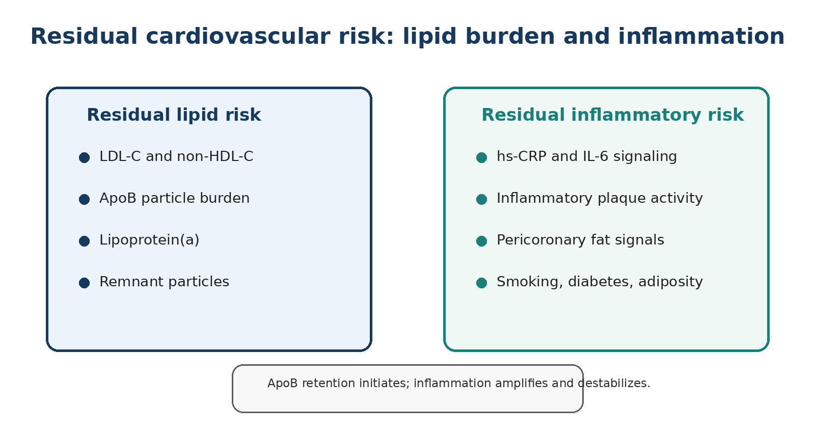
The modern model of atherosclerosis is not a contest between cholesterol and inflammation. ApoB-containing particles are required for common atherosclerosis to develop: they enter the intima, become retained and initiate a maladaptive immune response. Inflammation then amplifies lesion growth, impairs resolution, thins fibrous caps and can help convert a stable plaque into an event-producing plaque. A scientifically accurate shorthand is therefore: particle retention initiates; inflammation accelerates and destabilizes. [25-26,60-63]

Figure 1. Two interacting forms of residual cardiovascular risk. Original schematic for ElevatedCholesterol.com.
10.1 hs-CRP: useful context, not a diagnosis
High-sensitivity C-reactive protein (hs-CRP) is a reproducible marker of systemic inflammatory activity and a validated cardiovascular risk enhancer. It is not specific to coronary inflammation: infection, obesity, autoimmune disease, injury and many other conditions can raise it. A single elevated value should therefore be repeated when the patient is clinically well. Persistent hs-CRP elevation can identify residual inflammatory risk after LDL-C has been controlled, but it does not prove that a particular coronary plaque is inflamed.
Lp(a)-related vascular inflammation may exist even when hs-CRP is low. Lp(a) carries oxidized phospholipids and can activate monocytes and the arterial wall locally. Conversely, hs-CRP can be high in a person whose Lp(a) is low. The two markers answer different questions and should not be treated as interchangeable.
10.2 What the anti-inflammatory trials actually showed
CANTOS provided proof of principle that selectively reducing interleukin-1β signaling could reduce recurrent cardiovascular events without lowering LDL-C. The benefit was accompanied by more fatal infection and high cost, so canakinumab did not become routine atherosclerosis therapy. CIRT was equally instructive in the opposite direction: low-dose methotrexate did not lower IL-1β, IL-6 or hs-CRP and did not reduce events. Together, the trials showed that not every anti-inflammatory drug, and not every inflammatory pathway, is cardioprotective. [62-63]
COLCOT and LoDoCo2 tested low-dose colchicine in secondary prevention. COLCOT enrolled patients soon after myocardial infarction; LoDoCo2 enrolled patients with chronic coronary disease. LoDoCo2 reported a 31% relative reduction in its primary composite outcome with colchicine 0.5 mg daily. This is a relative—not absolute—risk reduction, and it applies to a selected secondary-prevention population, not to every person with elevated Lp(a) or CAC. [60-61]
More recent evidence makes the colchicine story less uniform. In CLEAR-SYNERGY, more than 7,000 patients with acute myocardial infarction undergoing PCI received colchicine or placebo, and colchicine did not significantly reduce cardiovascular death, recurrent myocardial infarction, stroke or ischemia-driven revascularization despite lowering CRP. Taken together, COLCOT and LoDoCo2 support cardiovascular benefit in selected settings, but the neutral CLEAR-SYNERGY result argues against treating colchicine as a universally beneficial therapy for all patients after myocardial infarction. [73]
| Trial | Population | Intervention | Main result | What it does not prove |
|---|---|---|---|---|
| CANTOS | Prior MI and hs-CRP ≥2 mg/L | Canakinumab | Reduced recurrent events without LDL-C lowering | That broad immune suppression is safe or practical for routine prevention |
| CIRT | Stable ASCVD plus diabetes/metabolic syndrome | Low-dose methotrexate | No reduction in inflammatory biomarkers or events | That inflammation is irrelevant; the targeted pathway was not effectively reduced |
| COLCOT | Recent myocardial infarction | Colchicine 0.5 mg/day | Lower composite ischemic events | Benefit in low-risk primary prevention |
| LoDoCo2 | Chronic coronary disease | Colchicine 0.5 mg/day | 31% relative reduction in primary composite outcome | Automatic treatment for high CAC or high Lp(a) |
Low-dose colchicine is an evidence-based option for selected patients with established coronary disease in jurisdictions where it is approved or guideline-supported. Renal or hepatic impairment, interacting drugs, gastrointestinal intolerance, myotoxicity risk and the uncertain non-cardiovascular mortality signal in LoDoCo2 require clinician oversight. It is not a supplement and should not be self-prescribed.
10.3 Imaging inflammation: pericoronary fat attenuation
CCTA-derived pericoronary fat attenuation attempts to capture biological signals emitted from an inflamed artery into the surrounding adipose tissue. CRISP-CT linked an abnormal fat attenuation index with future cardiac mortality independent of conventional CCTA findings. Commercial platforms can now produce vessel-level inflammatory metrics, but thresholds, scanner dependence, reproducibility and incremental clinical utility remain active areas of research. These tools are promising risk refiners, not replacements for symptoms, plaque burden, stenosis, ApoB or blood pressure. [20]
11. Can coronary plaque regress? Landmark imaging trials
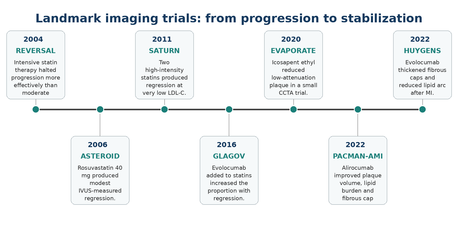
Plaque regression is real, but the phrase is frequently misunderstood. It usually means a modest reduction in measured plaque volume within an imaged arterial segment, not the disappearance of coronary disease. An intervention can also make plaque safer without shrinking it substantially: lipid pools may contract, fibrous caps may thicken, macrophage signals may fall and calcium density may rise. Clinical events remain the decisive outcome; imaging is a mechanistic bridge, not a substitute for adequately powered outcome trials. [55-56]

Figure 2. Selected imaging trials that shaped the modern concept of plaque regression and stabilization.
A series of landmark IVUS, OCT and CCTA trials — ASTEROID, SATURN, GLAGOV, PACMAN-AMI, HUYGENS and EVAPORATE — established that intensive LDL-lowering therapy can produce measurable regression and, more consistently, plaque stabilization: thicker fibrous caps, smaller lipid cores and less inflammation, even when overall volume change is modest. The key figures from each trial are summarized below; for the complete trial-by-trial breakdown, including REVERSAL, PRECISE-IVUS and the practical question of whether to repeat CAC or CCTA after starting therapy, see our complete plaque regression guide. [48-54]
Myth: plaque regression means that 40% of the plaque disappeared. Fact: GLAGOV showed an average PAV reduction of 0.95 percentage points and a greater proportion of patients with any measured regression. The artery did not become plaque-free.
| Trial | Imaging | Therapy | Key quantitative finding | Clinical lesson |
|---|---|---|---|---|
| ASTEROID | IVUS | Rosuvastatin 40 mg | PAV −0.98%; median TAV −6.8% | Intensive statin therapy can induce modest regression |
| GLAGOV | IVUS | Evolocumab + statin | PAV −0.95% vs +0.05%; regression in 64.3% vs 47.3% | Additional LDL lowering produces additional regression |
| PACMAN-AMI | IVUS + NIRS + OCT | Alirocumab + high-intensity statin | PAV −2.13% vs −0.92%; thicker caps and less lipid | Plaque volume and vulnerability can improve together |
| HUYGENS | OCT + IVUS | Evolocumab + statin | Fibrous cap +42.7 vs +21.5 µm; lipid arc fell more | Stabilization can be visible within one year after MI |
| EVAPORATE | CCTA | Icosapent ethyl 4 g/day | Low-attenuation plaque decreased in a small trial | Suggestive imaging mechanism; not proof for all fish-oil products |
Can Atherosclerotic Plaque Really Regress? — the complete guide, with full discussion of every landmark trial listed above.
Why CAC can rise while risk falls
Serial imaging studies show that statins can shift plaque toward denser calcification while reducing noncalcified plaque and clinical events. This does not mean that every increase in CAC is beneficial; it means that CAC progression is an unreliable treatment-response marker once lipid-lowering therapy has begun. The 2026 VitaK-CAC trial also illustrates the unresolved problem: slowing CAC progression may be biologically interesting, but no threshold of CAC change has been validated as a surrogate for fewer myocardial infarctions or deaths. [44-45,64-65]
12. Lifestyle, high-dose EPA and supplements: evidence vs hype
Lifestyle does not materially normalize genetically elevated Lp(a), but it changes the biological environment in which Lp(a) acts. Blood pressure, smoking, insulin resistance, sleep, visceral adiposity, cardiorespiratory fitness and dietary quality alter absolute risk. The strongest lifestyle message is not that one food or supplement dissolves plaque; it is that sustained risk-factor control lowers event probability over decades and can support favorable plaque biology when combined with proven medical therapy.
12.1 Physical activity and the 7,000-step claim
The statement that 7,000 steps per day produces '12.5% greater plaque reduction' comes from a small, retrospective, post hoc analysis of pooled trial data in patients recovering from acute coronary syndrome. Among 62 patients with analyzable IVUS, higher daily steps and an achieved LDL-C below 70 mg/dL were associated with greater plaque-volume reduction; the 7,000-step cut point was the cohort median, not a biologically proven threshold. The study is hypothesis-generating and cannot establish that moving from 6,900 to 7,100 steps causes a 12.5% plaque reduction. [66]
The practical conclusion remains strong but less sensational: regular activity should be prescribed because randomized cardiac-rehabilitation evidence reduces cardiovascular mortality and hospitalization, improves blood pressure and insulin sensitivity, and supports endothelial function. Step counts are useful adherence tools. The optimal target depends on baseline fitness, frailty, orthopedic limitations and progression over time; more activity is generally better than less until injury or excessive training risk becomes relevant.
12.2 Mediterranean-style eating patterns
PREDIMED showed fewer major cardiovascular events in high-risk primary-prevention participants assigned to Mediterranean diets supplemented with extra-virgin olive oil or nuts. Small imaging studies, including DISCO-CT, suggest that intensive dietary support added to optimal medical therapy may reduce noncalcified plaque more than medical therapy alone, but plaque-imaging trials are not large enough to isolate a single food as a regression therapy. [67-69]
For elevated Lp(a), the dietary goal is indirect: keep ApoB, blood pressure, glycemia and body composition favorable. A pattern centered on vegetables, legumes, whole grains as tolerated, nuts, extra-virgin olive oil, fish, minimally processed protein sources and limited trans fat is more defensible than a supplement stack marketed as a plaque remover.
12.3 High-dose EPA: prescription evidence is not the same as fish oil
REDUCE-IT randomized 8,179 statin-treated patients with established cardiovascular disease or diabetes plus risk factors, triglycerides 135-499 mg/dL and LDL-C 41-100 mg/dL to icosapent ethyl 2 g twice daily or placebo. The primary composite endpoint occurred in 17.2% versus 22.0% (hazard ratio 0.75). Atrial fibrillation or flutter hospitalization was more frequent, and serious bleeding showed a numerical increase. The result supports icosapent ethyl for selected high-risk patients meeting an evidence-based indication; it does not validate over-the-counter fish oil for plaque regression. [57]
EVAPORATE was a much smaller mechanistic CCTA trial. In statin-treated patients with elevated triglycerides and coronary atherosclerosis, icosapent ethyl reduced low-attenuation plaque volume compared with placebo over 18 months. The result is biologically coherent with REDUCE-IT, but the sample was small and imaging was not the clinical endpoint. By contrast, STRENGTH tested a high-dose EPA+DHA formulation and found no cardiovascular benefit. Formulation, population, comparator and achieved EPA exposure matter. [58-59]
12.4 Vitamin K2 after the 2026 VitaK-CAC trial
VitaK-CAC randomized 180 symptomatic adults with baseline CAC 50-400 Agatston units to menaquinone-7 (MK-7) 360 µg daily or placebo for two years. MK-7 significantly attenuated CAC progression. In the placebo group, median CAC rose from 145 to 214; in the active group it rose from 135 to 184. This is the first notable randomized signal that MK-7 may modify coronary calcification progression in mild coronary disease. [64]
The limitations are equally important. The trial was small, conducted at two Dutch centers, had dropouts, and measured a surrogate imaging endpoint rather than myocardial infarction, stroke or death. CAC progression is not a fully validated treatment-response surrogate, particularly because established therapies such as statins can increase calcification density while reducing events. The appropriate conclusion is 'promising and worthy of larger outcomes trials,' not 'K2 reverses heart disease.' Patients taking vitamin K antagonists require particular caution because vitamin K supplementation can interfere with anticoagulation.
12.5 Supplements promoted for plaque removal
Nattokinase, serrapeptase, rhamnan sulfate, intravenous phosphatidylcholine and similar protocols are frequently marketed using mechanistic language about fibrin, glycocalyx repair or lipid removal. None has evidence comparable to statins, ezetimibe, PCSK9 inhibition, blood-pressure treatment, smoking cessation or cardiac rehabilitation for preventing myocardial infarction and stroke. Small biomarker or uncontrolled imaging reports cannot establish plaque regression or net clinical benefit. Enzymes with fibrinolytic effects can also increase bleeding risk and are especially concerning around surgery or when combined with antiplatelet or anticoagulant drugs.
| Intervention | Evidence for events | Evidence for plaque imaging | Responsible interpretation |
|---|---|---|---|
| Mediterranean dietary pattern | Randomized outcome benefit in high-risk primary prevention | Small supportive imaging studies | Foundation of risk reduction; not a substitute for indicated medication |
| Regular aerobic + resistance activity | Strong rehabilitation and risk-factor evidence | Small IVUS/CCTA studies; 7,000-step result is post hoc | Aim for progressive, sustainable activity rather than a magic threshold |
| Icosapent ethyl 4 g/day | REDUCE-IT benefit in selected statin-treated high-risk patients | EVAPORATE low-attenuation plaque signal | Prescription therapy for an evidence-based indication, not generic fish oil |
| Vitamin K2 (MK-7) | No hard-outcome proof | VitaK-CAC slowed CAC progression | Promising but not standard plaque therapy |
| Berberine | No robust ASCVD outcome trial | No convincing coronary regression trial | May modestly affect lipids/glucose; not a statin replacement |
| Nattokinase / serrapeptase / Plaquex | No reliable outcome evidence | Insufficient or uncontrolled evidence | Do not present as proven plaque regression therapy |
13. One-time genetic therapies and the next decade
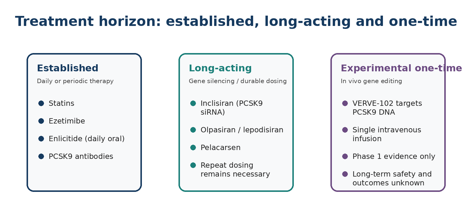
Cardiovascular therapeutics are moving from daily enzyme inhibition to infrequent gene silencing and, experimentally, permanent gene editing. These categories should not be conflated. Inclisiran and Lp(a)-directed siRNA reduce production of a target protein for months, but the biological effect eventually wanes and repeat dosing is required. Base editing changes a DNA letter in hepatocytes with the intention of creating a durable, possibly lifelong effect.

Figure 3. Established dosing, long-acting gene silencing and experimental one-time editing are distinct therapeutic categories.
13.1 VERVE-102: what the phase 1 evidence shows
VERVE-102 packages messenger RNA encoding an adenine base editor and a guide RNA targeting PCSK9 in a liver-directed lipid nanoparticle. In the open-label phase 1 Heart-2 study, 35 adults with heterozygous familial hypercholesterolemia or premature coronary disease received one infusion across six dose cohorts. Mean PCSK9 reductions ranged from 51% to 88%; mean LDL-C reductions ranged from 9% to 62%, with the largest effect at the highest dose. Reductions appeared durable during follow-up, which exceeded one year in 15 participants. Mild-to-moderate infusion reactions and transient alanine aminotransferase elevations occurred; the small study cannot define rare or long-term risks. [70]
The social-media phrase 'cuts LDL in half forever' is therefore premature. The highest dose produced a mean 62% LDL-C reduction in a small, nonrandomized dose-escalation study, not in a phase 3 outcomes trial. Durability beyond the current follow-up, off-target editing, immunogenicity, liver safety, reproductive implications, retreatment options and the consequences of permanently low PCSK9 all require longer observation. Phase 2 evaluation is the next step.
13.2 Enlicitide: a major advance, but not gene silencing
Enlicitide (Lipfendra) is an oral macrocyclic peptide that binds circulating PCSK9; it does not edit genes or silence PCSK9 mRNA. The CORALreef Lipids phase 3 trial reported approximately 56% placebo-adjusted LDL-C lowering on background therapy, with reductions in ApoB and Lp(a) as secondary lipid effects. The U.S. FDA approved Lipfendra in July 2026 as an adjunct to diet and exercise for adults with hypercholesterolemia, including heterozygous familial hypercholesterolemia. Cardiovascular outcomes data are still pending, so its approval is based on LDL-C lowering rather than proven event reduction with this specific agent. [71-72]
13.3 The ethical and clinical threshold for permanent editing
A daily drug can be stopped; an antibody wears off; an siRNA effect gradually recovers. A successful DNA edit is intentionally difficult to reverse. That raises the safety threshold. Early use is most likely to focus on people with severe inherited disease or premature ASCVD whose lifetime risk and treatment burden are high enough to justify uncertainty. Broad primary-prevention use would require exceptional evidence of editing specificity, long-term liver safety, durable event reduction, equitable access and informed consent.
VERVE-102 is investigational phase 1 therapy. Enlicitide is FDA-approved for LDL-C lowering but does not yet have completed cardiovascular outcomes evidence. Pelacarsen, olpasiran, lepodisiran, zerlasiran and muvalaplin remain investigational for Lp(a) lowering until outcomes and regulatory review establish their clinical role.
14. A practical clinical framework
The following framework is educational and must be individualized. It is designed to prevent two common errors: ignoring a high Lp(a) because no specific drug is approved, and overreacting to the biomarker with unproven tests or treatments.
Measure Lp(a) at least once in adulthood using the laboratory-reported unit. Do not use a fixed mg/dL-to-nmol/L conversion.
Confirm the clinical context: premature ASCVD, aortic stenosis, familial hypercholesterolemia, family history, kidney or liver disease, and whether the sample was obtained during major acute illness.
Measure the modifiable atherogenic burden: LDL-C, non-HDL-C and preferably ApoB, together with blood pressure, glycemia, smoking and body composition.
Estimate absolute risk. Use age-appropriate clinical tools, but recognize that calculators can underestimate lifetime genetic risk.
Use CAC selectively in asymptomatic adults when it can change the intensity of prevention. Interpret CAC zero as strongly reassuring for near-term risk, not as proof that Lp(a) is irrelevant.
Use CCTA for symptoms or a specific anatomical question, not as automatic screening for every elevated Lp(a) result.
Set LDL-C and ApoB goals according to global risk and plaque burden. High Lp(a) supports choosing a more intensive strategy when the decision is borderline.
Use proven therapy first: statin, ezetimibe, PCSK9-directed therapy, bempedoic acid or other approved treatments according to indication and tolerance.
Discuss aspirin only in the context of established ASCVD or an individualized primary-prevention assessment that explicitly weighs bleeding risk.
Offer family testing and revisit the plan when major outcome trials or guidelines change.
Before ordering any scan or adding any drug, ask: “What decision will change if this result is positive, negative or unchanged?” If no decision changes, the test may not be useful.
15. Clinical scenarios
Scenario A: high Lp(a), CAC zero
A 45-year-old nonsmoker has Lp(a) 130 mg/dL, LDL-C 118 mg/dL, normal blood pressure and no diabetes. CAC is zero. The calcium result is strongly reassuring for near-term risk, but the inherited Lp(a) level and decades of future exposure remain relevant.
A reasonable discussion would focus on family screening, maintaining very low lifetime exposure to smoking and hypertension, and whether LDL-C/ApoB lowering is appropriate given age, family history and preferences. Routine CCTA solely to search for soft plaque is not automatically required. A repeat CAC after an appropriate interval may be considered only if it would alter the prevention plan.
Scenario B: elevated Lp(a), CAC 208, minimal stenosis on CCTA
A 52-year-old has Lp(a) 138 mg/dL and CAC 208. CCTA shows a small calcified plaque with approximately 10% stenosis and no major left-main or LAD plaque. This is nonobstructive coronary atherosclerosis: the calcium score proves disease is present, while CCTA is reassuring that the current luminal narrowing is minimal.
The priority is not another scan. It is durable reduction of LDL-C and ApoB, blood pressure and metabolic risk, exercise, smoking avoidance, and shared decision-making about adding a PCSK9 inhibitor if the desired ApoB/LDL-C level is not achieved or overall risk warrants it. The high Lp(a) supports intensive prevention, but minimal stenosis does not mean zero risk and CAC 208 does not mean an intervention is required.
Scenario C: high Lp(a), symptoms and CAC zero
A 48-year-old with Lp(a) 160 mg/dL develops exertional chest pressure. A prior CAC score was zero. The symptom changes the question. CAC zero does not exclude noncalcified plaque or other causes of ischemic symptoms. Diagnostic evaluation may include CCTA or another appropriate test based on clinical assessment; repeating a calcium score alone would not answer the question.
Scenario D: high Lp(a), CAC 650 and prior myocardial infarction
A 64-year-old with prior myocardial infarction, Lp(a) 190 mg/dL and CAC 650 is a secondary-prevention patient with extensive disease. Treatment intensity is determined by established ASCVD, not by waiting for a dedicated Lp(a) drug. High-intensity or maximally tolerated statin therapy, ezetimibe and often a PCSK9 inhibitor are considered according to LDL-C/ApoB and local guidance. Aspirin or other antithrombotic therapy follows secondary-prevention indications. Clinical-trial eligibility may be relevant.
17. Key take-home messages
Lp(a) is an LDL-like ApoB particle with an additional apo(a) protein and oxidized-phospholipid cargo.
Its concentration is predominantly inherited and should usually be measured at least once in adult life.
Risk rises continuously; 50 mg/dL or approximately 105-125 nmol/L is a pragmatic risk-enhancing threshold, not a biological cliff.
Do not use a fixed formula to convert mg/dL and nmol/L.
Lp(a) is causal for ASCVD and calcific aortic stenosis, but absolute risk depends on the entire clinical profile.
CAC and Lp(a) are complementary. CAC zero is strongly reassuring for near-term risk; CAC at least 100 plus high Lp(a) identifies a substantially higher-risk group.
CAC cannot show noncalcified plaque or stenosis. CCTA can, but should answer a specific clinical question rather than be used as universal screening.
Serial CCTA studies link high Lp(a) with low-attenuation plaque progression, greater plaque burden and pericoronary inflammation.
Today, the central treatment is aggressive control of modifiable ApoB-containing particles and all other risk factors.
PCSK9 monoclonal antibodies provide strong LDL-C lowering and modest Lp(a) lowering; statins and ezetimibe remain foundational despite little direct Lp(a) effect.
Aspirin is not automatic in primary prevention; bleeding risk must be weighed carefully.
Pelacarsen, olpasiran, lepodisiran, zerlasiran and muvalaplin are investigational until cardiovascular outcomes and regulatory decisions establish their role.
High Lp(a) is important but not destiny. Earlier knowledge creates more time to prevent cumulative risk.
Glossary
| Term | Meaning |
|---|---|
| Apo(a) | Apolipoprotein(a), the Lp(a)-specific protein encoded by LPA. |
| ApoB | Apolipoprotein B; one molecule is present on each atherogenic particle, making it a marker of particle number. |
| ASCVD | Atherosclerotic cardiovascular disease, including coronary, cerebrovascular and peripheral arterial disease. |
| CAC | Coronary artery calcium, measured by noncontrast CT, usually reported as an Agatston score. |
| CCTA | Coronary computed tomography angiography, a contrast CT examination of coronary plaque, lumen and stenosis. |
| High-risk plaque | An imaging term for features associated with adverse outcomes, such as low attenuation or positive remodeling; not a guarantee of rupture. |
| hs-CRP | High-sensitivity C-reactive protein, a systemic inflammatory marker. |
| KIV-2 | Kringle IV type 2 repeat region in apo(a), a major genetic determinant of apo(a) size and Lp(a) concentration. |
| LDL-C | The estimated cholesterol mass carried within LDL particles. |
| Low-attenuation plaque | Very low-density plaque on CCTA, used as an imaging marker of lipid-rich or necrotic plaque. |
| Lp(a) | Lipoprotein(a), an LDL-like ApoB particle covalently linked to apo(a). |
| OxPL | Oxidized phospholipids, inflammatory lipids carried prominently by Lp(a). |
| PCSK9 | A protein that promotes LDL-receptor degradation; inhibiting it increases hepatic LDL clearance. |
| Percent atheroma volume | The proportion of the vessel volume occupied by plaque, used in quantitative imaging studies. |
Bibliography verification: journal/PubMed/official-source metadata checked 7 August 2026; independent clinical review remains pending.
References
1. Berg K. A new serum type system in man—the Lp system. Acta Pathol Microbiol Scand. 1963;59:369-382.
2. Reyes-Soffer G, Ginsberg HN, Berglund L, et al. Lipoprotein(a): a genetically determined, causal, and prevalent risk factor for atherosclerotic cardiovascular disease. Arterioscler Thromb Vasc Biol. 2022;42:e48-e60. doi:10.1161/ATV.0000000000000147.
3. Kronenberg F, Mora S, Stroes ESG, et al. Lipoprotein(a) in atherosclerotic cardiovascular disease and aortic stenosis: a European Atherosclerosis Society consensus statement. Eur Heart J. 2022;43(39):3925-3946. doi:10.1093/eurheartj/ehac361.
4. Koschinsky ML, Bajaj A, Boffa MB, et al. A focused update to the 2019 NLA scientific statement on use of lipoprotein(a) in clinical practice. J Clin Lipidol. 2024;18(3):e308-e319. doi:10.1016/j.jacl.2024.03.001.
5. Nordestgaard BG, Langsted A. Lipoprotein(a) and cardiovascular disease. Lancet. 2024;404:1952-1964.
6. Clarke R, Peden JF, Hopewell JC, et al. Genetic variants associated with Lp(a) lipoprotein level and coronary disease. N Engl J Med. 2009;361:2518-2528. doi:10.1056/NEJMoa0902604.
7. Kamstrup PR, Tybjærg-Hansen A, Steffensen R, Nordestgaard BG. Genetically elevated lipoprotein(a) and increased risk of myocardial infarction. JAMA. 2009;301:2331-2339.
8. Erqou S, Kaptoge S, Perry PL, et al. Lipoprotein(a) concentration and the risk of coronary heart disease, stroke, and nonvascular mortality. JAMA. 2009;302:412-423.
9. Björnson E, Adiels M, Taskinen MR, et al. Lipoprotein(a) is markedly more atherogenic than LDL: an apolipoprotein B-based genetic analysis. J Am Coll Cardiol. 2024;83:385-395. doi:10.1016/j.jacc.2023.10.039.
10. van der Valk FM, Bekkering S, Kroon J, et al. Oxidized phospholipids on lipoprotein(a) elicit arterial wall inflammation and an inflammatory monocyte response in humans. Circulation. 2016;134:611-624. doi:10.1161/CIRCULATIONAHA.116.020838.
11. Thanassoulis G, Campbell CY, Owens DS, et al. Genetic associations with valvular calcification and aortic stenosis. N Engl J Med. 2013;368:503-512. doi:10.1056/NEJMoa1109034.
12. Zheng KH, Tsimikas S, Pawade T, et al. Lipoprotein(a) and oxidized phospholipids promote valve calcification in patients with aortic stenosis. J Am Coll Cardiol. 2019;73:2150-2162.
13. Capoulade R, Chan KL, Yeang C, et al. Oxidized phospholipids, lipoprotein(a), and progression of calcific aortic valve stenosis. J Am Coll Cardiol. 2015;66:1236-1246.
14. Wong ND, Fan W, Hu X, et al. Lipoprotein(a) and Long-Term Cardiovascular Risk in a Multi-Ethnic Pooled Prospective Cohort. J Am Coll Cardiol. 2024;83(16):1511-1525. doi:10.1016/j.jacc.2024.02.031.
15. Mehta A, Vasquez N, Ayers CR, et al. Independent association of lipoprotein(a) and coronary artery calcification with atherosclerotic cardiovascular risk. J Am Coll Cardiol. 2022;79:757-768. doi:10.1016/j.jacc.2021.11.058.
16. Kaiser Y, Daghem M, Tzolos E, et al. Association of lipoprotein(a) with atherosclerotic plaque progression. J Am Coll Cardiol. 2022;79:223-233. doi:10.1016/j.jacc.2021.10.044.
17. Nurmohamed NS, Gaillard EL, Malkasian S, et al. Lipoprotein(a) and long-term plaque progression, low-density plaque, and pericoronary inflammation. JAMA Cardiol. 2024;9:826-834. doi:10.1001/jamacardio.2024.1874.
18. Chen X, Zheng Y, Lin S, et al. Lipoprotein(a) and plaque progression: insights from serial coronary computed tomography angiography and quantitative plaque assessment. Front Cardiovasc Med. 2026;13:1699503. doi:10.3389/fcvm.2026.1699503.
19. Cury RC, Leipsic J, Abbara S, et al. CAD-RADS 2.0—2022 Coronary Artery Disease Reporting and Data System. J Cardiovasc Comput Tomogr. 2022;16:536-557. doi:10.1016/j.jcct.2022.07.002.
20. Oikonomou EK, Marwan M, Desai MY, et al. Non-invasive detection of coronary inflammation using computed tomography and prediction of residual cardiovascular risk: the CRISP-CT study. Lancet. 2018;392:929-939. doi:10.1016/S0140-6736(18)31114-0.
21. Narula J, Stuckey TD, Nakazawa G, et al. Prospective deep learning-based quantitative assessment of coronary plaque by computed tomography angiography compared with intravascular ultrasound: the REVEALPLAQUE study. Eur Heart J Cardiovasc Imaging. 2024;25(9):1287-1295. doi:10.1093/ehjci/jeae115.
22. Dundas J, Leipsic J, Fairbairn T, et al. Interaction of AI-Enabled Quantitative Coronary Plaque Volumes on Coronary CT Angiography, FFRCT, and Clinical Outcomes: A Retrospective Analysis of the ADVANCE Registry. Circ Cardiovasc Imaging. 2024;17(3):e016143. doi:10.1161/CIRCIMAGING.123.016143.
23. Arnett DK, Blumenthal RS, Albert MA, et al. 2019 ACC/AHA guideline on the primary prevention of cardiovascular disease. Circulation. 2019;140:e596-e646.
24. Mach F, Koskinas KC, Roeters van Lennep JE, et al. 2025 Focused Update of the 2019 ESC/EAS Guidelines for the management of dyslipidaemias. Eur Heart J. 2025;46(42):4359-4378. doi:10.1093/eurheartj/ehaf190. Correction: Eur Heart J. 2026;47(6):697. doi:10.1093/eurheartj/ehaf1036.
25. Ference BA, Ginsberg HN, Graham I, et al. Low-density lipoproteins cause atherosclerotic cardiovascular disease: evidence from genetic, epidemiologic, and clinical studies. Eur Heart J. 2017;38:2459-2472.
26. Borén J, Chapman MJ, Krauss RM, et al. Low-density lipoproteins cause atherosclerotic cardiovascular disease: pathophysiological, genetic, and therapeutic insights. Eur Heart J. 2020;41:2313-2330.
27. O’Donoghue ML, Fazio S, Giugliano RP, et al. Lipoprotein(a), PCSK9 inhibition, and cardiovascular risk. Circulation. 2019;139:1483-1492.
28. Szarek M, Bittner VA, Aylward P, et al. Lipoprotein(a) lowering by alirocumab reduces the total burden of cardiovascular events independent of LDL-C lowering: ODYSSEY OUTCOMES. Eur Heart J. 2020;41:4245-4255.
29. Ray KK, Wright RS, Kallend D, et al. Two phase 3 trials of inclisiran in patients with elevated LDL cholesterol. N Engl J Med. 2020;382:1507-1519.
30. Nissen SE, Lincoff AM, Brennan D, et al. Bempedoic acid and cardiovascular outcomes in statin-intolerant patients. N Engl J Med. 2023;388:1353-1364.
31. Albers JJ, Slee A, O’Brien KD, et al. Relationship of apolipoproteins and lipoprotein(a) to cardiovascular outcomes: the AIM-HIGH trial. J Am Coll Cardiol. 2013;62:1575-1579.
32. Landray MJ, Haynes R, Hopewell JC, et al. Effects of extended-release niacin with laropiprant in high-risk patients. N Engl J Med. 2014;371:203-212.
33. Bhatia HS, Trainor P, Carlisle S, et al. Aspirin and Cardiovascular Risk in Individuals With Elevated Lipoprotein(a): The Multi-Ethnic Study of Atherosclerosis. J Am Heart Assoc. 2024;13(3):e033562. doi:10.1161/JAHA.123.033562.
34. Lacaze P, et al. Aspirin for primary prevention of cardiovascular events in relation to lipoprotein(a) genotypes. J Am Coll Cardiol. 2022;80:1287-1298.
35. Tsimikas S, Karwatowska-Prokopczuk E, Gouni-Berthold I, et al. Lipoprotein(a) reduction in persons with cardiovascular disease. N Engl J Med. 2020;382:244-255. doi:10.1056/NEJMoa1905239.
36. O’Donoghue ML, Rosenson RS, Gencer B, et al. Small interfering RNA to reduce lipoprotein(a) in cardiovascular disease. N Engl J Med. 2022;387:1855-1864. doi:10.1056/NEJMoa2211023.
37. Nissen SE, Ni W, Shen X, et al. Lepodisiran—a long-duration small interfering RNA targeting lipoprotein(a). N Engl J Med. 2025;392:1673-1683. doi:10.1056/NEJMoa2415818.
38. Nissen SE, Wolski K, Watts GF, et al. Single ascending and multiple-dose trial of zerlasiran, a short interfering RNA targeting lipoprotein(a). JAMA. 2024;331:1534-1543. doi:10.1001/jama.2024.4504.
39. Nissen SE, Wang Q, Nicholls SJ, et al. Zerlasiran—a small-interfering RNA targeting lipoprotein(a): a phase 2 randomized clinical trial. JAMA. 2024;332:1992-2002. doi:10.1001/jama.2024.21957.
40. Nicholls SJ, Ni W, Rhodes GM, et al. Oral muvalaplin for lowering of lipoprotein(a): a randomized clinical trial. JAMA. 2025;333:222-231. doi:10.1001/jama.2024.24017.
41. ClinicalTrials.gov. NCT04023552. Lp(a)HORIZON: assessing the impact of lipoprotein(a) lowering with pelacarsen on major cardiovascular events. Record accessed 20 July 2026.
42. ClinicalTrials.gov. NCT05581303. OCEAN(a)-Outcomes: olpasiran trials of cardiovascular events and lipoprotein(a) reduction. Record accessed 20 July 2026.
43. ClinicalTrials.gov. NCT06292013. ACCLAIM-Lp(a): lepodisiran and major adverse cardiovascular events. Record accessed 20 July 2026.
44. Puri R, Nicholls SJ, Shao M, et al. Impact of statins on serial coronary calcification during atheroma progression and regression. J Am Coll Cardiol. 2015;65:1273-1282.
45. Lee SE, Chang HJ, Sung JM, et al. Effects of statins on coronary atherosclerotic plaques: the PARADIGM study. JACC Cardiovasc Imaging. 2018;11:1475-1484.
46. Cegla J, Neely RDG, France M, et al. HEART UK consensus statement on lipoprotein(a): a call to action. Atherosclerosis. 2019;291:62-70. doi:10.1016/j.atherosclerosis.2019.10.011.
47. O'Donoghue ML, Rosenson RS, Lopez JAG, et al; OCEAN(a)-DOSE Trial Investigators. The Off-Treatment Effects of Olpasiran on Lipoprotein(a) Lowering: OCEAN(a)-DOSE Extension Period Results. J Am Coll Cardiol. 2024;84(9):790-797. doi:10.1016/j.jacc.2024.05.058.
48. Nissen SE, Nicholls SJ, Sipahi I, et al. Effect of very high-intensity statin therapy on regression of coronary atherosclerosis: the ASTEROID trial. JAMA. 2006;295:1556-1565. doi:10.1001/jama.295.13.jpc60002.
49. Nicholls SJ, Ballantyne CM, Barter PJ, et al. Effect of two intensive statin regimens on progression of coronary disease. N Engl J Med. 2011;365:2078-2087. doi:10.1056/NEJMoa1110874.
50. Nicholls SJ, Puri R, Anderson T, et al. Effect of evolocumab on progression of coronary disease in statin-treated patients: the GLAGOV randomized clinical trial. JAMA. 2016;316:2373-2384. doi:10.1001/jama.2016.16951.
51. Sabatine MS, Giugliano RP, Keech AC, et al. Evolocumab and clinical outcomes in patients with cardiovascular disease. N Engl J Med. 2017;376:1713-1722. doi:10.1056/NEJMoa1615664.
52. Schwartz GG, Steg PG, Szarek M, et al. Alirocumab and cardiovascular outcomes after acute coronary syndrome. N Engl J Med. 2018;379:2097-2107. doi:10.1056/NEJMoa1801174.
53. Räber L, Ueki Y, Otsuka T, et al. Effect of alirocumab added to high-intensity statin therapy on coronary atherosclerosis in patients with acute myocardial infarction: the PACMAN-AMI randomized clinical trial. JAMA. 2022;327:1771-1781. doi:10.1001/jama.2022.5218.
54. Nicholls SJ, Kataoka Y, Nissen SE, et al. Effect of evolocumab on coronary plaque phenotype and burden in statin-treated patients following myocardial infarction. JACC Cardiovasc Imaging. 2022;15:1308-1321. doi:10.1016/j.jcmg.2022.03.002.
55. Puri R, et al. Coronary atherosclerotic plaque regression: JACC state-of-the-art review. J Am Coll Cardiol. 2022;79:66-82. doi:10.1016/j.jacc.2021.10.035.
56. Mancini GBJ, et al. Atherosclerotic coronary plaque regression and risk of adverse cardiovascular events: a systematic review and updated meta-regression analysis. JAMA Cardiol. 2023;8:937-946. doi:10.1001/jamacardio.2023.2731.
57. Bhatt DL, Steg PG, Miller M, et al. Cardiovascular risk reduction with icosapent ethyl for hypertriglyceridemia. N Engl J Med. 2019;380:11-22. doi:10.1056/NEJMoa1812792.
58. Budoff MJ, Bhatt DL, Kinninger A, et al. Effect of icosapent ethyl on progression of coronary atherosclerosis in patients with elevated triglycerides on statin therapy: final results of the EVAPORATE trial. Eur Heart J. 2020;41:3925-3932. doi:10.1093/eurheartj/ehaa652.
59. Nicholls SJ, Lincoff AM, Garcia M, et al. Effect of high-dose omega-3 fatty acids vs corn oil on major adverse cardiovascular events in patients at high cardiovascular risk: the STRENGTH randomized clinical trial. JAMA. 2020;324:2268-2280. doi:10.1001/jama.2020.22258.
60. Nidorf SM, Fiolet ATL, Mosterd A, et al. Colchicine in patients with chronic coronary disease. N Engl J Med. 2020;383:1838-1847. doi:10.1056/NEJMoa2021372.
61. Tardif JC, Kouz S, Waters DD, et al. Efficacy and safety of low-dose colchicine after myocardial infarction. N Engl J Med. 2019;381:2497-2505. doi:10.1056/NEJMoa1912388.
62. Ridker PM, Everett BM, Thuren T, et al. Antiinflammatory therapy with canakinumab for atherosclerotic disease. N Engl J Med. 2017;377:1119-1131. doi:10.1056/NEJMoa1707914.
63. Ridker PM, Everett BM, Pradhan A, et al. Low-dose methotrexate for the prevention of atherosclerotic events. N Engl J Med. 2019;380:752-762. doi:10.1056/NEJMoa1809798.
64. Vossen LM, de Leeuw PW, Schurgers LJ, et al. Two years of menaquinone-7 supplementation and coronary artery calcification: a randomized clinical trial. JAMA Cardiol. Published online June 10, 2026. doi:10.1001/jamacardio.2026.1279.
65. Blaha MJ, Choi S. Coronary artery calcium progression—A useful outcome in clinical trials? JAMA Cardiol. Published online June 10, 2026. doi:10.1001/jamacardio.2026.1267.
66. Nishitani-Yokoyama M, et al. Preliminary pilot study of combined effects of physical activity and achievement of LDL-cholesterol target on coronary plaque volume changes in patients with acute coronary syndrome. J Clin Med. 2020;9:1578. doi:10.3390/jcm9051578.
67. Smit JM, et al. High-risk coronary plaque regression after intensive lifestyle intervention in nonobstructive coronary disease: a randomized study. JACC Cardiovasc Imaging. 2021;14:1192-1202. doi:10.1016/j.jcmg.2020.10.019.
68. Estruch R, Ros E, Salas-Salvadó J, et al. Primary prevention of cardiovascular disease with a Mediterranean diet supplemented with extra-virgin olive oil or nuts. N Engl J Med. 2018;378:e34. doi:10.1056/NEJMoa1800389.
69. Madssen E, Moholdt T, Videm V, et al. Coronary atheroma regression and plaque characteristics assessed by grayscale and radiofrequency intravascular ultrasound after aerobic exercise. Am J Cardiol. 2014;114:1504-1511. doi:10.1016/j.amjcard.2014.08.012.
70. Vafai SB, Täubel J, Ashdown T, et al. In vivo base editing of PCSK9 with VERVE-102 for hypercholesterolemia. N Engl J Med. Published online May 25, 2026. doi:10.1056/NEJMoa2601283.
71. Navar AM, Mikhailova E, Catapano AL, et al. A placebo-controlled trial of the oral PCSK9 inhibitor enlicitide. N Engl J Med. 2026;394:529-539. doi:10.1056/NEJMoa2511002.
72. U.S. Food and Drug Administration. FDA approves first oral PCSK9 inhibitor to lower LDL cholesterol in adults with high cholesterol. July 17, 2026.
73. Jolly SS, d'Entremont MA, Lee SF, et al; CLEAR Investigators. Colchicine in Acute Myocardial Infarction. N Engl J Med. 2025;392(7):633-642. doi:10.1056/NEJMoa2405922.
16. Frequently asked questions and common myths
Can diet lower Lp(a)?
Usually not by a clinically important amount. Diet can lower LDL-C, blood pressure, weight and diabetes risk, which still reduces the total risk associated with high Lp(a).
Can exercise lower Lp(a)?
Regular exercise generally has little effect on the Lp(a) number, but improves fitness, blood pressure, insulin sensitivity, body composition and overall cardiovascular risk.
Does a normal LDL-C cancel high Lp(a)?
No. Lp(a) is an independent risk factor. However, a low LDL-C and ApoB reduce the modifiable background risk and are among the most effective actions available today.
Is ApoB more important than Lp(a)?
They answer different questions. ApoB measures total atherogenic particle number; Lp(a) identifies a genetically distinct and particularly atherogenic subset. Both may matter.
Does CAC zero mean there is no plaque?
No. It means no detectable calcified plaque. Noncalcified plaque can exist, especially in younger or symptomatic people. In asymptomatic adults, CAC zero is nevertheless strongly reassuring for near-term risk.
Should everyone with high Lp(a) have CCTA?
No. CCTA is most useful for symptoms or a specific anatomical question that can change management. Universal screening has not been shown to improve outcomes.
Can plaque regress?
Total and noncalcified plaque can stabilize or regress with intensive risk-factor treatment, while calcified volume may increase. The clinically important goal is fewer events and a more stable plaque phenotype, not a perfect scan.
Does a statin increase coronary calcium?
It can increase calcified plaque or calcium density while reducing lipid-rich plaque and events. This is one reason serial CAC is not a simple treatment-response test.
Does vitamin K2 remove coronary calcium?
No convincing randomized evidence shows that vitamin K2 removes established coronary plaque or reduces cardiovascular events. It should not replace proven lipid and blood-pressure treatment.
Should I take aspirin because Lp(a) is high?
Not automatically. Selected high-Lp(a) groups may benefit, but primary-prevention aspirin can cause serious bleeding. The decision requires individualized medical assessment.
Do PCSK9 inhibitors treat Lp(a)?
They are approved LDL-lowering drugs that also lower Lp(a) modestly. Their event-reduction evidence cannot be attributed solely to Lp(a) lowering.
Is very low LDL-C dangerous?
Randomized PCSK9 trials have achieved very low LDL-C without a clear increase in neurocognitive events, hemorrhagic stroke or other major safety outcomes over trial follow-up. Targets should still be individualized.
Should Lp(a) be repeated?
Usually one adult measurement is enough. Repeating can be reasonable if the first result may have been affected by major illness, kidney or liver dysfunction, or a laboratory-method concern.
Can Lp(a) explain aortic stenosis?
It is a causal risk factor for calcific aortic valve disease, but not the only cause. High Lp(a) does not prove that a murmur or valve abnormality is due to Lp(a).
When will specific Lp(a) drugs be available?
That depends on outcomes, regulatory review, safety, manufacturing and reimbursement. As of July 2026, no dedicated Lp(a)-lowering outcomes result had established routine therapy.
“My Lp(a) is genetic, so nothing I do matters.”
Genetics determines much of the Lp(a) concentration, but it does
not
determine blood pressure, smoking, diabetes, LDL-C/ApoB exposure,
fitness, adherence or whether proven treatment is used early.
Glossary
| Term | Meaning |
|---|---|
| Apo(a) | Apolipoprotein(a), the Lp(a)-specific protein encoded by LPA. |
| ApoB | Apolipoprotein B; one molecule is present on each atherogenic particle, making it a marker of particle number. |
| ASCVD | Atherosclerotic cardiovascular disease, including coronary, cerebrovascular and peripheral arterial disease. |
| CAC | Coronary artery calcium, measured by noncontrast CT, usually reported as an Agatston score. |
| CCTA | Coronary computed tomography angiography, a contrast CT examination of coronary plaque, lumen and stenosis. |
| High-risk plaque | An imaging term for features associated with adverse outcomes, such as low attenuation or positive remodeling; not a guarantee of rupture. |
| hs-CRP | High-sensitivity C-reactive protein, a systemic inflammatory marker. |
| KIV-2 | Kringle IV type 2 repeat region in apo(a), a major genetic determinant of apo(a) size and Lp(a) concentration. |
| LDL-C | The estimated cholesterol mass carried within LDL particles. |
| Low-attenuation plaque | Very low-density plaque on CCTA, used as an imaging marker of lipid-rich or necrotic plaque. |
| Lp(a) | Lipoprotein(a), an LDL-like ApoB particle covalently linked to apo(a). |
| OxPL | Oxidized phospholipids, inflammatory lipids carried prominently by Lp(a). |
| PCSK9 | A protein that promotes LDL-receptor degradation; inhibiting it increases hepatic LDL clearance. |
| Percent atheroma volume | The proportion of the vessel volume occupied by plaque, used in quantitative imaging studies. |
Bibliography verification: journal/PubMed/official-source metadata checked 7 August 2026; independent clinical review remains pending.
References
1. Berg K. A new serum type system in man—the Lp system. Acta Pathol Microbiol Scand. 1963;59:369-382.
2. Reyes-Soffer G, Ginsberg HN, Berglund L, et al. Lipoprotein(a): a genetically determined, causal, and prevalent risk factor for atherosclerotic cardiovascular disease. Arterioscler Thromb Vasc Biol. 2022;42:e48-e60. doi:10.1161/ATV.0000000000000147.
3. Kronenberg F, Mora S, Stroes ESG, et al. Lipoprotein(a) in atherosclerotic cardiovascular disease and aortic stenosis: a European Atherosclerosis Society consensus statement. Eur Heart J. 2022;43(39):3925-3946. doi:10.1093/eurheartj/ehac361.
4. Koschinsky ML, Bajaj A, Boffa MB, et al. A focused update to the 2019 NLA scientific statement on use of lipoprotein(a) in clinical practice. J Clin Lipidol. 2024;18(3):e308-e319. doi:10.1016/j.jacl.2024.03.001.
5. Nordestgaard BG, Langsted A. Lipoprotein(a) and cardiovascular disease. Lancet. 2024;404:1952-1964.
6. Clarke R, Peden JF, Hopewell JC, et al. Genetic variants associated with Lp(a) lipoprotein level and coronary disease. N Engl J Med. 2009;361:2518-2528. doi:10.1056/NEJMoa0902604.
7. Kamstrup PR, Tybjærg-Hansen A, Steffensen R, Nordestgaard BG. Genetically elevated lipoprotein(a) and increased risk of myocardial infarction. JAMA. 2009;301:2331-2339.
8. Erqou S, Kaptoge S, Perry PL, et al. Lipoprotein(a) concentration and the risk of coronary heart disease, stroke, and nonvascular mortality. JAMA. 2009;302:412-423.
9. Björnson E, Adiels M, Taskinen MR, et al. Lipoprotein(a) is markedly more atherogenic than LDL: an apolipoprotein B-based genetic analysis. J Am Coll Cardiol. 2024;83:385-395. doi:10.1016/j.jacc.2023.10.039.
10. van der Valk FM, Bekkering S, Kroon J, et al. Oxidized phospholipids on lipoprotein(a) elicit arterial wall inflammation and an inflammatory monocyte response in humans. Circulation. 2016;134:611-624. doi:10.1161/CIRCULATIONAHA.116.020838.
11. Thanassoulis G, Campbell CY, Owens DS, et al. Genetic associations with valvular calcification and aortic stenosis. N Engl J Med. 2013;368:503-512. doi:10.1056/NEJMoa1109034.
12. Zheng KH, Tsimikas S, Pawade T, et al. Lipoprotein(a) and oxidized phospholipids promote valve calcification in patients with aortic stenosis. J Am Coll Cardiol. 2019;73:2150-2162.
13. Capoulade R, Chan KL, Yeang C, et al. Oxidized phospholipids, lipoprotein(a), and progression of calcific aortic valve stenosis. J Am Coll Cardiol. 2015;66:1236-1246.
14. Wong ND, Fan W, Hu X, et al. Lipoprotein(a) and Long-Term Cardiovascular Risk in a Multi-Ethnic Pooled Prospective Cohort. J Am Coll Cardiol. 2024;83(16):1511-1525. doi:10.1016/j.jacc.2024.02.031.
15. Mehta A, Vasquez N, Ayers CR, et al. Independent association of lipoprotein(a) and coronary artery calcification with atherosclerotic cardiovascular risk. J Am Coll Cardiol. 2022;79:757-768. doi:10.1016/j.jacc.2021.11.058.
16. Kaiser Y, Daghem M, Tzolos E, et al. Association of lipoprotein(a) with atherosclerotic plaque progression. J Am Coll Cardiol. 2022;79:223-233. doi:10.1016/j.jacc.2021.10.044.
17. Nurmohamed NS, Gaillard EL, Malkasian S, et al. Lipoprotein(a) and long-term plaque progression, low-density plaque, and pericoronary inflammation. JAMA Cardiol. 2024;9:826-834. doi:10.1001/jamacardio.2024.1874.
18. Chen X, Zheng Y, Lin S, et al. Lipoprotein(a) and plaque progression: insights from serial coronary computed tomography angiography and quantitative plaque assessment. Front Cardiovasc Med. 2026;13:1699503. doi:10.3389/fcvm.2026.1699503.
19. Cury RC, Leipsic J, Abbara S, et al. CAD-RADS 2.0—2022 Coronary Artery Disease Reporting and Data System. J Cardiovasc Comput Tomogr. 2022;16:536-557. doi:10.1016/j.jcct.2022.07.002.
20. Oikonomou EK, Marwan M, Desai MY, et al. Non-invasive detection of coronary inflammation using computed tomography and prediction of residual cardiovascular risk: the CRISP-CT study. Lancet. 2018;392:929-939. doi:10.1016/S0140-6736(18)31114-0.
21. Narula J, Stuckey TD, Nakazawa G, et al. Prospective deep learning-based quantitative assessment of coronary plaque by computed tomography angiography compared with intravascular ultrasound: the REVEALPLAQUE study. Eur Heart J Cardiovasc Imaging. 2024;25(9):1287-1295. doi:10.1093/ehjci/jeae115.
22. Dundas J, Leipsic J, Fairbairn T, et al. Interaction of AI-Enabled Quantitative Coronary Plaque Volumes on Coronary CT Angiography, FFRCT, and Clinical Outcomes: A Retrospective Analysis of the ADVANCE Registry. Circ Cardiovasc Imaging. 2024;17(3):e016143. doi:10.1161/CIRCIMAGING.123.016143.
23. Arnett DK, Blumenthal RS, Albert MA, et al. 2019 ACC/AHA guideline on the primary prevention of cardiovascular disease. Circulation. 2019;140:e596-e646.
24. Mach F, Koskinas KC, Roeters van Lennep JE, et al. 2025 Focused Update of the 2019 ESC/EAS Guidelines for the management of dyslipidaemias. Eur Heart J. 2025;46(42):4359-4378. doi:10.1093/eurheartj/ehaf190. Correction: Eur Heart J. 2026;47(6):697. doi:10.1093/eurheartj/ehaf1036.
25. Ference BA, Ginsberg HN, Graham I, et al. Low-density lipoproteins cause atherosclerotic cardiovascular disease: evidence from genetic, epidemiologic, and clinical studies. Eur Heart J. 2017;38:2459-2472.
26. Borén J, Chapman MJ, Krauss RM, et al. Low-density lipoproteins cause atherosclerotic cardiovascular disease: pathophysiological, genetic, and therapeutic insights. Eur Heart J. 2020;41:2313-2330.
27. O’Donoghue ML, Fazio S, Giugliano RP, et al. Lipoprotein(a), PCSK9 inhibition, and cardiovascular risk. Circulation. 2019;139:1483-1492.
28. Szarek M, Bittner VA, Aylward P, et al. Lipoprotein(a) lowering by alirocumab reduces the total burden of cardiovascular events independent of LDL-C lowering: ODYSSEY OUTCOMES. Eur Heart J. 2020;41:4245-4255.
29. Ray KK, Wright RS, Kallend D, et al. Two phase 3 trials of inclisiran in patients with elevated LDL cholesterol. N Engl J Med. 2020;382:1507-1519.
30. Nissen SE, Lincoff AM, Brennan D, et al. Bempedoic acid and cardiovascular outcomes in statin-intolerant patients. N Engl J Med. 2023;388:1353-1364.
31. Albers JJ, Slee A, O’Brien KD, et al. Relationship of apolipoproteins and lipoprotein(a) to cardiovascular outcomes: the AIM-HIGH trial. J Am Coll Cardiol. 2013;62:1575-1579.
32. Landray MJ, Haynes R, Hopewell JC, et al. Effects of extended-release niacin with laropiprant in high-risk patients. N Engl J Med. 2014;371:203-212.
33. Bhatia HS, Trainor P, Carlisle S, et al. Aspirin and Cardiovascular Risk in Individuals With Elevated Lipoprotein(a): The Multi-Ethnic Study of Atherosclerosis. J Am Heart Assoc. 2024;13(3):e033562. doi:10.1161/JAHA.123.033562.
34. Lacaze P, et al. Aspirin for primary prevention of cardiovascular events in relation to lipoprotein(a) genotypes. J Am Coll Cardiol. 2022;80:1287-1298.
35. Tsimikas S, Karwatowska-Prokopczuk E, Gouni-Berthold I, et al. Lipoprotein(a) reduction in persons with cardiovascular disease. N Engl J Med. 2020;382:244-255. doi:10.1056/NEJMoa1905239.
36. O’Donoghue ML, Rosenson RS, Gencer B, et al. Small interfering RNA to reduce lipoprotein(a) in cardiovascular disease. N Engl J Med. 2022;387:1855-1864. doi:10.1056/NEJMoa2211023.
37. Nissen SE, Ni W, Shen X, et al. Lepodisiran—a long-duration small interfering RNA targeting lipoprotein(a). N Engl J Med. 2025;392:1673-1683. doi:10.1056/NEJMoa2415818.
38. Nissen SE, Wolski K, Watts GF, et al. Single ascending and multiple-dose trial of zerlasiran, a short interfering RNA targeting lipoprotein(a). JAMA. 2024;331:1534-1543. doi:10.1001/jama.2024.4504.
39. Nissen SE, Wang Q, Nicholls SJ, et al. Zerlasiran—a small-interfering RNA targeting lipoprotein(a): a phase 2 randomized clinical trial. JAMA. 2024;332:1992-2002. doi:10.1001/jama.2024.21957.
40. Nicholls SJ, Ni W, Rhodes GM, et al. Oral muvalaplin for lowering of lipoprotein(a): a randomized clinical trial. JAMA. 2025;333:222-231. doi:10.1001/jama.2024.24017.
41. ClinicalTrials.gov. NCT04023552. Lp(a)HORIZON: assessing the impact of lipoprotein(a) lowering with pelacarsen on major cardiovascular events. Record accessed 20 July 2026.
42. ClinicalTrials.gov. NCT05581303. OCEAN(a)-Outcomes: olpasiran trials of cardiovascular events and lipoprotein(a) reduction. Record accessed 20 July 2026.
43. ClinicalTrials.gov. NCT06292013. ACCLAIM-Lp(a): lepodisiran and major adverse cardiovascular events. Record accessed 20 July 2026.
44. Puri R, Nicholls SJ, Shao M, et al. Impact of statins on serial coronary calcification during atheroma progression and regression. J Am Coll Cardiol. 2015;65:1273-1282.
45. Lee SE, Chang HJ, Sung JM, et al. Effects of statins on coronary atherosclerotic plaques: the PARADIGM study. JACC Cardiovasc Imaging. 2018;11:1475-1484.
46. Cegla J, Neely RDG, France M, et al. HEART UK consensus statement on lipoprotein(a): a call to action. Atherosclerosis. 2019;291:62-70. doi:10.1016/j.atherosclerosis.2019.10.011.
47. O'Donoghue ML, Rosenson RS, Lopez JAG, et al; OCEAN(a)-DOSE Trial Investigators. The Off-Treatment Effects of Olpasiran on Lipoprotein(a) Lowering: OCEAN(a)-DOSE Extension Period Results. J Am Coll Cardiol. 2024;84(9):790-797. doi:10.1016/j.jacc.2024.05.058.
48. Nissen SE, Nicholls SJ, Sipahi I, et al. Effect of very high-intensity statin therapy on regression of coronary atherosclerosis: the ASTEROID trial. JAMA. 2006;295:1556-1565. doi:10.1001/jama.295.13.jpc60002.
49. Nicholls SJ, Ballantyne CM, Barter PJ, et al. Effect of two intensive statin regimens on progression of coronary disease. N Engl J Med. 2011;365:2078-2087. doi:10.1056/NEJMoa1110874.
50. Nicholls SJ, Puri R, Anderson T, et al. Effect of evolocumab on progression of coronary disease in statin-treated patients: the GLAGOV randomized clinical trial. JAMA. 2016;316:2373-2384. doi:10.1001/jama.2016.16951.
51. Sabatine MS, Giugliano RP, Keech AC, et al. Evolocumab and clinical outcomes in patients with cardiovascular disease. N Engl J Med. 2017;376:1713-1722. doi:10.1056/NEJMoa1615664.
52. Schwartz GG, Steg PG, Szarek M, et al. Alirocumab and cardiovascular outcomes after acute coronary syndrome. N Engl J Med. 2018;379:2097-2107. doi:10.1056/NEJMoa1801174.
53. Räber L, Ueki Y, Otsuka T, et al. Effect of alirocumab added to high-intensity statin therapy on coronary atherosclerosis in patients with acute myocardial infarction: the PACMAN-AMI randomized clinical trial. JAMA. 2022;327:1771-1781. doi:10.1001/jama.2022.5218.
54. Nicholls SJ, Kataoka Y, Nissen SE, et al. Effect of evolocumab on coronary plaque phenotype and burden in statin-treated patients following myocardial infarction. JACC Cardiovasc Imaging. 2022;15:1308-1321. doi:10.1016/j.jcmg.2022.03.002.
55. Puri R, et al. Coronary atherosclerotic plaque regression: JACC state-of-the-art review. J Am Coll Cardiol. 2022;79:66-82. doi:10.1016/j.jacc.2021.10.035.
56. Mancini GBJ, et al. Atherosclerotic coronary plaque regression and risk of adverse cardiovascular events: a systematic review and updated meta-regression analysis. JAMA Cardiol. 2023;8:937-946. doi:10.1001/jamacardio.2023.2731.
57. Bhatt DL, Steg PG, Miller M, et al. Cardiovascular risk reduction with icosapent ethyl for hypertriglyceridemia. N Engl J Med. 2019;380:11-22. doi:10.1056/NEJMoa1812792.
58. Budoff MJ, Bhatt DL, Kinninger A, et al. Effect of icosapent ethyl on progression of coronary atherosclerosis in patients with elevated triglycerides on statin therapy: final results of the EVAPORATE trial. Eur Heart J. 2020;41:3925-3932. doi:10.1093/eurheartj/ehaa652.
59. Nicholls SJ, Lincoff AM, Garcia M, et al. Effect of high-dose omega-3 fatty acids vs corn oil on major adverse cardiovascular events in patients at high cardiovascular risk: the STRENGTH randomized clinical trial. JAMA. 2020;324:2268-2280. doi:10.1001/jama.2020.22258.
60. Nidorf SM, Fiolet ATL, Mosterd A, et al. Colchicine in patients with chronic coronary disease. N Engl J Med. 2020;383:1838-1847. doi:10.1056/NEJMoa2021372.
61. Tardif JC, Kouz S, Waters DD, et al. Efficacy and safety of low-dose colchicine after myocardial infarction. N Engl J Med. 2019;381:2497-2505. doi:10.1056/NEJMoa1912388.
62. Ridker PM, Everett BM, Thuren T, et al. Antiinflammatory therapy with canakinumab for atherosclerotic disease. N Engl J Med. 2017;377:1119-1131. doi:10.1056/NEJMoa1707914.
63. Ridker PM, Everett BM, Pradhan A, et al. Low-dose methotrexate for the prevention of atherosclerotic events. N Engl J Med. 2019;380:752-762. doi:10.1056/NEJMoa1809798.
64. Vossen LM, de Leeuw PW, Schurgers LJ, et al. Two years of menaquinone-7 supplementation and coronary artery calcification: a randomized clinical trial. JAMA Cardiol. Published online June 10, 2026. doi:10.1001/jamacardio.2026.1279.
65. Blaha MJ, Choi S. Coronary artery calcium progression—A useful outcome in clinical trials? JAMA Cardiol. Published online June 10, 2026. doi:10.1001/jamacardio.2026.1267.
66. Nishitani-Yokoyama M, et al. Preliminary pilot study of combined effects of physical activity and achievement of LDL-cholesterol target on coronary plaque volume changes in patients with acute coronary syndrome. J Clin Med. 2020;9:1578. doi:10.3390/jcm9051578.
67. Smit JM, et al. High-risk coronary plaque regression after intensive lifestyle intervention in nonobstructive coronary disease: a randomized study. JACC Cardiovasc Imaging. 2021;14:1192-1202. doi:10.1016/j.jcmg.2020.10.019.
68. Estruch R, Ros E, Salas-Salvadó J, et al. Primary prevention of cardiovascular disease with a Mediterranean diet supplemented with extra-virgin olive oil or nuts. N Engl J Med. 2018;378:e34. doi:10.1056/NEJMoa1800389.
69. Madssen E, Moholdt T, Videm V, et al. Coronary atheroma regression and plaque characteristics assessed by grayscale and radiofrequency intravascular ultrasound after aerobic exercise. Am J Cardiol. 2014;114:1504-1511. doi:10.1016/j.amjcard.2014.08.012.
70. Vafai SB, Täubel J, Ashdown T, et al. In vivo base editing of PCSK9 with VERVE-102 for hypercholesterolemia. N Engl J Med. Published online May 25, 2026. doi:10.1056/NEJMoa2601283.
71. Navar AM, Mikhailova E, Catapano AL, et al. A placebo-controlled trial of the oral PCSK9 inhibitor enlicitide. N Engl J Med. 2026;394:529-539. doi:10.1056/NEJMoa2511002.
72. U.S. Food and Drug Administration. FDA approves first oral PCSK9 inhibitor to lower LDL cholesterol in adults with high cholesterol. July 17, 2026.
73. Jolly SS, d'Entremont MA, Lee SF, et al; CLEAR Investigators. Colchicine in Acute Myocardial Infarction. N Engl J Med. 2025;392(7):633-642. doi:10.1056/NEJMoa2405922.