
Executive Summary
Cardiovascular tests do not measure the same thing. A coronary artery calcium scan counts calcified coronary atherosclerosis on a noncontrast CT. Coronary CT angiography, or CCTA, uses contrast to visualize the coronary lumen and can identify both calcified and noncalcified plaque. Blood markers such as LDL-C, ApoB, lipoprotein(a), triglycerides and hsCRP do not image plaque at all, but they describe biological drivers and residual risk that imaging cannot replace.
The 2026 ACC/AHA dyslipidemia guideline places selective CAC use inside a broader primary-prevention framework. For appropriate adults with borderline or intermediate PREVENT-ASCVD risk, CAC can reclassify uncertainty and help set the intensity of prevention. A positive CAC score proves that coronary atherosclerosis is already present, but a zero score does not prove that every coronary artery is plaque-free because noncalcified plaque can exist before calcium develops.
CCTA answers a different question. It can identify stenosis, plaque location and plaque composition, and it has a central role in selected patients with stable or acute chest pain who are not low risk. Modern reporting systems such as CAD-RADS 2.0 also recognize overall plaque burden and high-risk plaque features, not just the single tightest narrowing. This is important because many myocardial infarctions arise from plaques that were not severely obstructive beforehand.
Newer CCTA-derived tools are promising but should be placed in the correct tier. Perivascular fat attenuation index, including FAI-based approaches studied in CRISP-CT and ORFAN, can capture an inflammatory signal around coronary arteries and has prognostic value. Quantitative and AI-assisted plaque analysis can make plaque burden more reproducible. Neither should be marketed as a crystal ball that predicts the exact plaque or exact date of a heart attack.
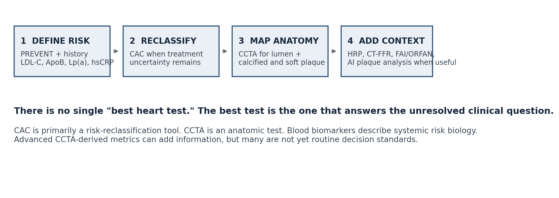
The practical rule is simple: start with the clinical question. Use a risk calculator and blood markers to define baseline risk, CAC when risk reclassification will change a prevention decision, CCTA when coronary anatomy is the unresolved question, and advanced add-ons only when they contribute information that is actionable in the specific clinical setting.
Related guides: CAC vs CCTA • coronary calcium score guide • high-risk coronary plaque on CCTA • preventive cardiology blood tests
1. The most important distinction: risk, plaque, stenosis and inflammation are different targets
A recurring diagnostic mistake is to treat every cardiovascular number as if it measures the same disease. It does not. LDL-C and ApoB estimate exposure to circulating atherogenic particles. Lp(a) captures an inherited risk pathway. hsCRP can reflect systemic inflammatory activity. CAC measures the burden of calcified coronary plaque. CCTA maps the coronary arteries and can show both plaque and stenosis. FAI-based analysis attempts to extract a local inflammatory signal from the fat surrounding coronary arteries.
This distinction matters because a normal result in one domain cannot automatically cancel an abnormal result in another. A person can have low short-term calculated risk but a strong family history or very high Lp(a). A CAC score of zero can coexist with noncalcified plaque. A CCTA can show substantial nonobstructive plaque even when there is no severe stenosis. Conversely, an isolated biomarker abnormality does not tell you how much plaque is already present.
| Clinical question | Best starting tool | What it answers | What it does not answer |
|---|---|---|---|
| What is my estimated future ASCVD risk? | PREVENT + clinical history | 10-year and, in selected ages, 30-year estimated risk | Whether plaque is already present in a specific artery |
| Is calcified coronary plaque already present? | CAC | Calcified plaque burden and risk reclassification | Soft plaque, exact stenosis, ischemia |
| What do my coronary arteries look like? | CCTA | Lumen, stenosis, plaque location and composition | Whether every lesion will cause a future event |
| What is driving residual risk? | Blood biomarkers | ApoB, Lp(a), TG/remnants, hsCRP and metabolic context | Direct plaque anatomy |
| Is there an inflammatory CT signal? | FAI / ORFAN-type analysis | Perivascular inflammatory signal and prognostic information | A fully standardized treatment target in routine practice |
2. Coronary artery calcium: the best-established CT risk reclassifier
CAC is obtained from a noncontrast, ECG-gated CT and reported most commonly as an Agatston score. The test is fast, does not require iodinated contrast and exposes the patient to a relatively low radiation dose with contemporary protocols. Its strongest role is not to diagnose chest pain or to map stenosis. Its strongest role is to improve risk classification when a preventive treatment decision is uncertain.
The 2026 ACC/AHA dyslipidemia guideline recommends selective CAC use in appropriate primary-prevention adults, particularly men age 40 and older and women age 45 and older with borderline or intermediate 10-year risk when the result would change management. Both the absolute CAC burden and its age- and sex-standardized percentile add prognostic information. Any CAC means coronary atherosclerosis is present, while progressively higher burdens generally support more intensive risk-factor treatment.
The trap is to overread a score of zero. CAC 0 is powerful negative risk information for many adults, but it does not mean "zero atherosclerosis" in a literal anatomical sense. Noncalcified plaque can exist before it becomes detectable as calcium, especially in younger people and in selected high-risk phenotypes. CAC also does not show whether a coronary segment is 20%, 50% or 80% narrowed.
Read: Coronary Calcium Score Explained: 0, 10, 100, 200, 400, 1000. For the imaging decision itself, see CAC vs CCTA: What Each Test Can and Cannot Tell You.
3. CCTA: when anatomy is the question
CCTA is contrast-enhanced CT imaging designed to visualize the coronary arteries. Unlike CAC, it can show the vessel lumen and characterize calcified and noncalcified plaque. This makes it useful when the unresolved question is anatomical: Is coronary disease present? Where is it? How much plaque is there? Is a lesion obstructive? Are there high-risk plaque characteristics?
The 2021 AHA/ACC chest pain guideline supports CCTA as a useful diagnostic and risk-stratification test in selected patients with stable chest pain at intermediate to high pretest risk and no known coronary disease. It is also one of the anatomic options for selected intermediate-risk acute chest pain patients. Low-risk patients should not automatically be sent for imaging simply because the technology exists. Testing is useful when the answer is likely to change the next clinical step.
CCTA is not perfect. Dense calcium can create blooming artifact and make stenosis appear worse. Image quality can fall with very high heart rates, arrhythmia, motion, obesity or suboptimal breath holding. Iodinated contrast introduces kidney and allergy considerations. A visually moderate stenosis also does not automatically prove flow-limiting ischemia, which is why CT fractional flow reserve, stress imaging or invasive physiology can be appropriate in selected cases.
Read: When Is CCTA Actually Worth Doing? and CAC vs CCTA: What Each Test Can and Cannot Tell You.
4. High-risk plaque: useful biology, not a countdown clock
Modern CCTA interpretation looks beyond the narrowest lumen. CAD-RADS 2.0 incorporates plaque burden and recognizes high-risk plaque features. Common features include low-attenuation plaque, positive remodeling, the napkin-ring sign and spotty calcification. In CAD-RADS, the high-risk plaque modifier is used when at least two qualifying high-risk features are present.
These findings matter because plaque biology and total plaque burden carry prognostic information beyond stenosis severity. A mildly narrowing plaque can still be biologically important. But the phrase "high-risk plaque" is easy to misuse. It does not mean that a radiologist can identify the one plaque that will definitely rupture next month. It is a population-level risk marker and should be integrated with total plaque burden, symptoms, risk factors and preventive treatment.
This distinction is one reason the site keeps a separate owner page for high-risk plaque. The hub should orient the reader, while the satellite article can explain attenuation thresholds, remodeling, CAD-RADS notation and the evidence in detail without forcing every reader through the same technical depth.
Read: High-Risk Coronary Plaque on CCTA.
5. Soft plaque vs calcified plaque: why the labels can mislead
"Soft plaque" is a popular shorthand for noncalcified plaque, but it is not a synonym for "about to rupture." CCTA can separate broad plaque components based on attenuation and can identify low-attenuation features associated with higher risk, but histology is more complicated than a binary soft-versus-hard model. Calcification also has two meanings that can appear contradictory: a higher total coronary calcium burden is associated with higher long-term risk, while increasing calcification within an individual treated plaque can be part of a stabilizing transformation.
Serial CCTA studies such as PARADIGM help explain the apparent paradox. Statin-treated plaques showed slower overall and noncalcified plaque progression, fewer high-risk features and more calcified plaque. That is why a rising calcium score after statin initiation cannot be interpreted simplistically as proof that treatment is failing. The clinical goal is fewer cardiovascular events and more stable plaque biology, not necessarily a lower Agatston number on every subsequent scan.
Read: Soft Plaque vs Calcified Plaque. For treatment-related change, see Can Atherosclerotic Plaque Really Regress? and Plaque Stabilization vs Regression.
6. Coronary inflammation on CT: FAI and ORFAN are promising, but not a universal routine test
Inflammation is central to atherosclerosis, but conventional CCTA does not directly image inflammatory cells. Perivascular fat attenuation index, or FAI, uses changes in the CT appearance of fat around coronary arteries as an indirect signal of vascular inflammation. CRISP-CT showed that perivascular FAI added prognostic information for mortality. The larger ORFAN program later showed that inflammatory risk derived from routine CCTA can further stratify patients, including many without obstructive coronary disease.
The signal is clinically interesting because nonobstructive disease is common and events can occur despite the absence of a severe narrowing. However, FAI-based risk tools should not be presented as if they are already equivalent to CAC or standard CCTA interpretation in every health system. Availability, software implementation, thresholds, regulatory status and how the result changes therapy are still evolving. In 2026 it is best viewed as an advanced CCTA-derived risk layer, not a mandatory test for every preventive cardiology patient.
Read: Coronary Inflammation: FAI / ORFAN.
7. AI plaque analysis: measurement can improve before outcomes evidence does
Artificial intelligence and semi-automated plaque software can quantify total plaque volume, noncalcified plaque, calcified plaque and other compositional features more consistently than a purely visual read. That can be valuable for research, longitudinal analysis and selected clinical programs. It can also reduce reader variability and make plaque burden more explicit in the report.
The marketing problem begins when improved measurement is presented as proven improved outcomes. A tool can be excellent at segmentation without having randomized evidence that using its output changes therapy in a way that prevents more myocardial infarctions. Different vendors also use different reconstruction methods, thresholds and plaque definitions. The correct question is not "Does this software use AI?" but "What does it measure, is it validated, is the result reproducible, and what decision will change because of it?"
Read: AI Plaque Analysis on CCTA: Real vs Marketing.
8. Aortic valve calcium: a different disease question
Aortic valve calcium is not simply another way to score coronary plaque. CT calcium quantification of the aortic valve can help when echocardiographic assessment of aortic stenosis is discordant or difficult to interpret, particularly in selected low-flow or low-gradient scenarios. The anatomical target, thresholds and clinical decision are different from coronary CAC.
This distinction is especially important in people with elevated Lp(a), because Lp(a) is associated not only with atherosclerotic cardiovascular disease but also with calcific aortic valve disease. A coronary calcium score and an aortic valve calcium score should therefore never be treated as interchangeable numbers.
Read: Aortic Valve Calcium Explained.
9. Blood tests are part of advanced diagnostics, even though they do not image plaque
Imaging shows disease burden and anatomy. Blood tests show exposure and biology. A strong preventive assessment usually needs both domains rather than forcing one to replace the other. LDL-C remains the familiar lipid measure, but ApoB can better represent atherogenic particle number in discordant or metabolically complex cases. Lp(a) identifies a largely inherited risk pathway and should be measured at least once in adulthood according to the 2026 ACC/AHA guideline. Triglycerides and non-HDL cholesterol help characterize remnant risk, while hsCRP can identify residual inflammatory risk in selected patients.
Kidney and metabolic markers also matter because the cardiovascular system does not operate in isolation. Glycemia or HbA1c, eGFR and urine albumin-to-creatinine ratio can materially change risk classification and treatment priorities. The point is not to order every biomarker marketed to consumers. It is to choose validated tests that can change a real clinical decision.
Read: Preventive Cardiology Blood Tests (2026). For risk integration, see PREVENT-ASCVD Calculator Explained: 10-Year vs 30-Year Risk.
10. Which test is usually the better next question?
| Scenario | Usually more useful | Why |
|---|---|---|
| Asymptomatic adult, prevention decision still uncertain after risk calculation | CAC | Best-established imaging tool for risk reclassification when the result will change preventive therapy. |
| Stable chest pain, intermediate to high pretest likelihood, no known CAD | CCTA or appropriate functional testing | Anatomy or ischemia is the unresolved question, not just long-term risk. |
| CAC 0 but major risk enhancer such as very high Lp(a) or strong family history | Clinical integration; sometimes CCTA if there is a separate anatomical indication | CAC 0 lowers risk but does not erase inherited risk or exclude all noncalcified plaque. |
| Known plaque on CCTA, want to understand morphology | CCTA interpretation with CAD-RADS / plaque features | Shows plaque distribution, stenosis and selected high-risk features. |
| Question is whether a moderate CCTA lesion causes ischemia | CT-FFR, stress imaging or invasive physiology in selected cases | Anatomical stenosis and physiological significance are related but not identical. |
| Known or suspected aortic stenosis with discordant echo findings | Aortic valve calcium CT in selected cases | Targets valve calcification, not coronary atherosclerosis. |
| Question is inherited or residual risk biology | Lp(a), ApoB, hsCRP and targeted laboratory testing | Blood biomarkers answer a different question from CT anatomy. |
Before requesting a test, write down the decision you are trying to make. If the answer will not change what you do, the test may add information without adding value. If the decision is prevention intensity, start with risk calculation and validated biomarkers. If uncertainty remains, consider whether CAC would reclassify it. If symptoms or coronary anatomy are the question, discuss whether CCTA or functional testing is the better tool.
11. Seven common interpretation errors
• "CAC 0 means I have no plaque." False. It means no detectable calcified coronary plaque on that scan. Noncalcified plaque can still exist.
• "CCTA is a better CAC scan." Wrong question. CCTA and CAC have different indications, acquisition methods and clinical roles.
• "A 30% stenosis is harmless." Nonobstructive plaque still contributes to future risk, and plaque burden matters beyond the single tightest lesion.
• "High-risk plaque means a heart attack is imminent." It is a prognostic feature, not a countdown clock or lesion-specific certainty.
• "More calcium after starting a statin proves plaque is worsening." Plaque calcification can increase as plaque phenotype becomes more stable.
• "AI plaque analysis is automatically superior to expert CCTA interpretation." Quantification can improve consistency, but clinical utility depends on validation and actionability.
• "One impressive test overrides every other risk signal." Imaging, biomarkers, symptoms, family history and treatment response must be integrated.
12. A sensible 2026 testing sequence
For prevention in an asymptomatic adult, the sequence usually begins with history, blood pressure, smoking status, diabetes and kidney status, a standard lipid profile, and targeted biomarkers such as Lp(a) and ApoB when appropriate. PREVENT-ASCVD then provides a contemporary estimate for adults in its intended age range. If that leaves a genuine treatment decision unresolved, CAC can reclassify risk in the appropriate population.
CCTA should not be added automatically after CAC. It enters when there is a separate reason to define coronary anatomy, most commonly symptoms or a specific clinical question that CAC cannot answer. Once CCTA exists, plaque burden, stenosis, high-risk features and selected advanced analyses can be extracted from that dataset. The value is in using more information from a clinically justified scan, not in generating serial scans simply to watch numbers move.
The same restraint applies to repeat testing. There is no evidence-based reason to repeat every test every year. The correct interval depends on what changed, whether the result can alter management, the baseline findings, symptoms and the specific test. A stable prevention plan with well-controlled risk factors often benefits more from adherence and periodic laboratory monitoring than from repeated radiation-based imaging.
13. FAQ
Is CAC or CCTA better for screening?
For an asymptomatic primary-prevention decision, CAC has the more established role as a selective risk-reclassification test. CCTA is an anatomical test and is generally used when anatomy is clinically relevant rather than as routine screening for everyone.
Can CCTA see soft plaque if my calcium score is zero?
Yes. CCTA can identify noncalcified plaque that a calcium scan does not detect. Whether CCTA should be performed is a separate clinical decision; the ability to see more does not mean every person with CAC 0 needs CCTA.
Does a high CAC score tell me which artery is dangerously | | blocked?
No. CAC quantifies calcified plaque burden but does not reliably define the degree of luminal stenosis. CCTA or other anatomical testing is needed when stenosis is the question.
Should I repeat CCTA to prove plaque regression?
Usually not as a routine self-tracking strategy. Serial CCTA has research value and selected clinical uses, but repeated contrast and radiation imaging should have a clear clinical indication.
Are FAI and AI plaque analysis ready for everyone?
No. Both are advancing rapidly and can add prognostic or quantitative information in selected settings, but access, standardization, evidence of management impact and integration into routine care remain uneven.
What if my imaging and blood tests disagree?
That is common because they measure different domains. For example, low current ApoB after treatment does not erase plaque accumulated over prior decades, and CAC 0 does not erase a genetically high Lp(a). The disagreement often adds information rather than proving one test is wrong.
References
1. Blumenthal RS, Morris PB, Gaudino M, et al. 2026 ACC/AHA/AACVPR/ABC/ACPM/ADA/AGS/APhA/ASPC/NLA/PCNA Guideline on the Management of Dyslipidemia. Circulation. 2026;153(17):e1154-e1276. doi:10.1161/CIR.0000000000001423.
2. Gulati M, Levy PD, Mukherjee D, et al. 2021 AHA/ACC/ASE/CHEST/SAEM/SCCT/SCMR Guideline for the Evaluation and Diagnosis of Chest Pain. Circulation. 2021;144(22):e368-e454. doi:10.1161/CIR.0000000000001029.
3. Narula J, Chandrashekhar Y, Ahmadi A, et al. SCCT 2021 Expert Consensus Document on Coronary Computed Tomographic Angiography. J Cardiovasc Comput Tomogr. 2021;15(3):192-217. doi:10.1016/j.jcct.2020.11.001.
4. Cury RC, Leipsic J, Abbara S, et al. CAD-RADS 2.0 - 2022 Coronary Artery Disease-Reporting and Data System. JACC Cardiovasc Imaging. 2022;15(11):1974-2001. doi:10.1016/j.jcmg.2022.07.002.
5. Shaw LJ, Blankstein R, Bax JJ, et al. Society of Cardiovascular Computed Tomography / North American Society of Cardiovascular Imaging Expert Consensus Document on Coronary CT Imaging of Atherosclerotic Plaque. J Cardiovasc Comput Tomogr. 2021;15(2):93-109. doi:10.1016/j.jcct.2020.11.002.
6. Newby DE, Adamson PD, Berry C, et al; SCOT-HEART Investigators. Coronary CT Angiography and 5-Year Risk of Myocardial Infarction. N Engl J Med. 2018;379(10):924-933. doi:10.1056/NEJMoa1805971.
7. Lee SE, Chang HJ, Sung JM, et al. Effects of Statins on Coronary Atherosclerotic Plaques: The PARADIGM Study. JACC Cardiovasc Imaging. 2018;11(10):1475-1484. doi:10.1016/j.jcmg.2018.04.015.
8. Oikonomou EK, Marwan M, Desai MY, et al. Non-invasive detection of coronary inflammation using computed tomography and prediction of residual cardiovascular risk: the CRISP-CT study. Lancet. 2018;392:929-939. doi:10.1016/S0140-6736(18)31114-0.
9. Chan K, Wahome E, Tsiachristas A, et al. Inflammatory risk and cardiovascular events in patients without obstructive coronary artery disease: the ORFAN multicentre, longitudinal cohort study. Lancet. 2024;403:2606-2618. doi:10.1016/S0140-6736(24)00596-8.
10. Koschinsky ML, Bajaj A, Boffa MB, et al. A focused update to the 2019 NLA scientific statement on use of lipoprotein(a) in clinical practice. J Clin Lipidol. 2024;18(3):e308-e319. doi:10.1016/j.jacl.2024.03.001.
11. Kronenberg F, Mora S, Stroes ESG, et al. Lipoprotein(a) in atherosclerotic cardiovascular disease and aortic stenosis: a European Atherosclerosis Society consensus statement. Eur Heart J. 2022;43(39):3925-3946. doi:10.1093/eurheartj/ehac361.