Executive Summary
Coronary CT angiography (CCTA) is uniquely positioned between a calcium scan and invasive angiography. It shows the lumen, but it also visualizes the arterial wall and can distinguish calcified, non-calcified and mixed plaque. Modern quantitative software can estimate total plaque volume, percent atheroma volume and plaque composition.
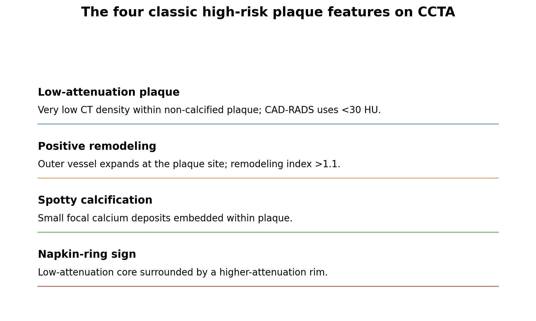
CAD-RADS 2.0 defines four classic CCTA high-risk plaque features: low-attenuation plaque (<30 Hounsfield units), positive remodeling (remodeling index >1.1), spotty calcification and the napkin-ring sign. The HRP modifier is used when a plaque clearly demonstrates at least two of these features.
High-risk plaque features predict events beyond stenosis severity, but their positive predictive value is modest because they are relatively common and most individual plaques do not cause an acute coronary syndrome. Contemporary imaging literature therefore emphasizes the combination of plaque phenotype, total plaque burden and stenosis rather than a binary “vulnerable plaque” label.
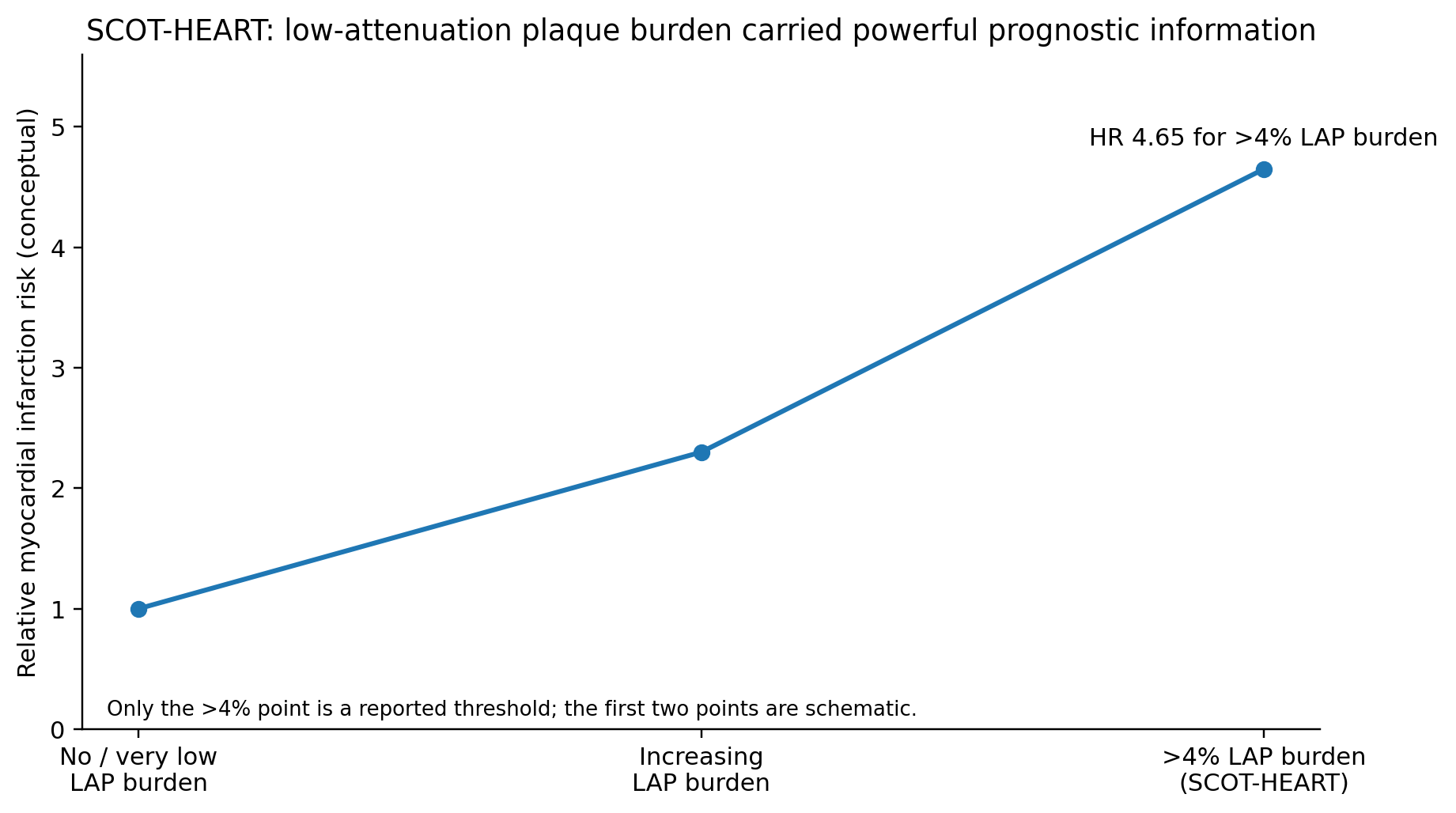
In SCOT-HEART, quantitative low-attenuation non-calcified plaque burden was the strongest predictor of subsequent fatal or nonfatal myocardial infarction. Patients with low-attenuation plaque burden >4% had an approximately 4.65-fold higher risk of myocardial infarction. This was a post hoc analysis in patients with stable chest pain, not a universal treatment threshold.
PROMISE similarly showed that the presence of high-risk plaque features was associated with a higher rate of major adverse cardiovascular events even after accounting for significant stenosis and conventional risk factors.
In 2025, the American College of Cardiology issued a scientific statement on quantitative coronary plaque analysis (QCPA), acknowledging growing accuracy and prognostic evidence and multiple FDA-cleared commercial tools, while emphasizing that evidence remains insufficient to prove that routine QCPA improves clinical outcomes or to establish one universal approach to serial plaque imaging.
The clinical response to high-risk plaque is usually aggressive preventive therapy: lower LDL-C/ApoB according to risk, control blood pressure and diabetes, eliminate smoking, address inflammation and lifestyle, and evaluate symptoms appropriately. Revascularization is primarily driven by anatomy plus ischemia/symptoms — not by a high-risk plaque label alone.
1. Why "Percent Blockage" Is an Incomplete Description of Coronary Disease
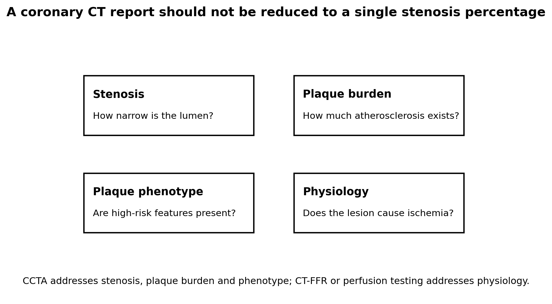
Atherosclerosis begins in the arterial wall, not in the lumen. Early plaques can grow outward through positive remodeling, preserving the lumen while the plaque burden increases. A report that says “25% stenosis” can therefore describe anything from a tiny stable plaque to a large outward-remodeled plaque with a lipid-rich core.
This explains a paradox that often confuses patients: many myocardial infarctions arise from lesions that were not severely obstructive before they ruptured. Severe stenosis matters, but the biology of the plaque and the total amount of disease matter too.
CCTA is valuable because it can show these dimensions noninvasively. The test should not be reduced to a binary answer of “blocked” versus “not blocked.”
2. The Four Classic High-Risk Plaque Features
| Feature | Common CCTA definition | Biological interpretation / limitation |
|---|---|---|
| Low-attenuation plaque | Non-calcified plaque containing focal attenuation <30 HU in CAD-RADS | Correlates with lipid-rich/necrotic material, but HU measurements vary with scanner, contrast and reconstruction. |
| Positive remodeling | Outer vessel diameter at plaque site >1.1× reference diameter | Suggests outward vessel expansion around a growing plaque; not specific for imminent rupture. |
| Spotty calcification | Small punctate calcium within plaque | Associated with active plaque biology; definitions vary by size and method. |
| Napkin-ring sign | Low-attenuation central plaque adjacent to lumen with a higher-attenuation peripheral rim | Specific high-risk morphology, but uncommon and observer dependent. |
3. Low-Attenuation Plaque: The Most Important Quantitative Signal So Far
Low-attenuation plaque (LAP) is attractive because CT density provides a quantitative window into plaque composition. Very low attenuation is associated with lipid-rich and necrotic-core components that are common in advanced atherosclerotic lesions.
The strongest long-term CCTA evidence comes from SCOT-HEART. Among 1,769 patients with stable chest pain followed for a median 4.7 years, low-attenuation plaque burden was the strongest predictor of fatal or nonfatal myocardial infarction after adjustment for risk score, CAC and coronary stenosis. The adjusted hazard ratio was 1.60 per doubling of LAP burden.
Patients with LAP burden >4% were nearly five times more likely to experience myocardial infarction (HR 4.65; 95% CI 2.06-10.5). The threshold is clinically memorable, but it should not be treated as a universal “danger line.” It was derived from a post hoc analysis in a specific symptomatic cohort, and software methods continue to evolve.
“More low-attenuation plaque predicts higher risk” is well supported. “Any plaque under 30 HU will rupture” is false. Risk applies probabilistically across a patient and coronary tree, not deterministically to one voxel or lesion.
4. Positive Remodeling: Large Plaque Can Hide Behind a Small Stenosis
Positive remodeling is the arterial wall’s outward expansion around plaque. It helps explain why the lumen can look only mildly narrowed while the vessel wall contains substantial atherosclerosis.
CAD-RADS 2.0 uses a remodeling index above 1.1 as a high-risk feature. Positive remodeling is associated with advanced plaque and acute coronary syndromes, but it should not be read as evidence that rupture is imminent.
Clinically, the key lesson is that “non-obstructive” does not mean “non-atherosclerotic” or “low lifetime risk.” A large non-obstructive plaque burden may warrant aggressive prevention even when no lesion is a revascularization target.
5. Spotty Calcium vs Dense Calcium
Coronary calcification is not one biological state. Small focal calcium deposits within otherwise non-calcified plaque — spotty calcification — have been associated with active and potentially unstable plaque biology. Dense sheet-like macrocalcification is more often seen in chronic, healed or stabilized plaque.
This distinction helps reconcile two apparently conflicting facts: a high calcium score indicates high total atherosclerotic burden and higher risk, while increased calcium density within an individual plaque during intensive statin treatment can be a feature of stabilization.
Therefore, neither “calcium is bad” nor “calcium is good” is sufficiently accurate. Pattern, burden, plaque context and treatment history matter.
6. The Napkin-Ring Sign
The napkin-ring sign describes a cross-sectional non-calcified plaque with a central low-attenuation region in contact with the lumen and a peripheral rim of higher attenuation. It is considered a high-risk feature because it correlates with advanced fibroatheroma morphology.
It is relatively uncommon and requires good image quality and experienced interpretation. Its presence increases concern, but absence does not make a plaque harmless and presence does not guarantee an event.
7. What PROMISE and SCOT-HEART Taught Us About Prognosis
| Study | Key finding | Practical lesson |
|---|---|---|
| PROMISE high-risk plaque analysis | High-risk plaque associated with higher MACE; reported event rate 6.4% vs 2.4% and HR ~2.7 in published analyses | High-risk morphology adds prognostic information beyond stenosis and clinical risk. |
| SCOT-HEART adverse plaque analysis | Adverse plaque features associated with coronary events, but relationship attenuated after accounting for CAC/plaque burden | Plaque phenotype matters, but total burden strongly influences risk. |
| SCOT-HEART quantitative LAP, 2020 | LAP burden strongest predictor of MI; >4% burden HR 4.65 | Quantitative composition can refine risk beyond stenosis and CAC. |
| RAPID-CTCA plaque analysis | Total, noncalcified and LAP burden predicted events more strongly than obstructive CAD in suspected ACS | Plaque burden can outperform a simple obstructive/non-obstructive label. |
8. Plaque Burden May Matter More Than "Vulnerability"
The original “vulnerable plaque” concept imagined that finding one dangerous lesion could identify the future culprit. Long-term imaging and pathology have made the picture more complex. Plaques evolve; high-risk features can appear and disappear; multiple lesions can coexist; thrombogenicity and systemic inflammation also influence whether rupture becomes an infarction.
A 2025 JACC: Cardiovascular Imaging position statement emphasizes that plaque volume/burden, morphology and stenosis all contribute to risk, and that plaque burden often emerges as the single strongest predictor of instability and events.
This changes the clinical target from “find and fix the one bad plaque” toward “treat the entire atherosclerotic disease process.”
The most dangerous finding on CCTA may not be the tightest stenosis. It may be a large overall burden of non-calcified and low-attenuation plaque distributed through the coronary tree.
9. CAD-RADS 2.0: How High-Risk Plaque Is Actually Reported
CAD-RADS 2.0 standardized CCTA reporting by separating stenosis severity, plaque burden and modifiers. Plaque burden is categorized from P1 (mild) to P4 (extensive), and the HRP modifier is added when a plaque clearly demonstrates two or more of the four high-risk features.
The system deliberately avoids calling a lesion “vulnerable plaque,” because the term can imply a certainty that CCTA cannot provide. “High-risk plaque” is more accurate: it describes a phenotype associated with higher probability of future events.
| Dimension | Example CAD-RADS concept |
|---|---|
| Stenosis | CAD-RADS 1–5 based on maximal luminal narrowing |
| Overall plaque burden | P1 mild, P2 moderate, P3 severe, P4 extensive |
| High-risk plaque | HRP modifier when ≥2 classic HRP features are clearly present |
| Ischemia | I+, I− or I± if CT-FFR or CT perfusion is performed |
| Context modifiers | Stent, graft, non-diagnostic segment, exceptions |
10. High-Risk Plaque Does Not Automatically Mean Stent
PCI treats a focal obstruction or culprit lesion; it does not cure the systemic plaque biology that created the disease. In stable coronary disease, revascularization decisions are generally driven by symptoms, severity/location of stenosis, documented ischemia and specific high-risk anatomy — not simply by the presence of low attenuation or positive remodeling.
Preventive therapy is therefore the first response to most non-obstructive high-risk plaque findings. This can include intensive LDL-C/ApoB lowering, blood-pressure control, diabetes treatment, smoking cessation, exercise, diet and other therapies according to the patient’s overall risk.
If a plaque is associated with a significant stenosis, symptoms or lesion-specific ischemia, the management pathway changes. CT-FFR, stress imaging or invasive physiology can determine whether the lesion is functionally important.
11. AI and Quantitative Coronary Plaque Analysis in 2026
Artificial intelligence has made whole-coronary-tree plaque quantification practical. Commercial systems can segment coronary vessels and quantify total, non-calcified, calcified and low-attenuation plaque, sometimes within minutes rather than hours of expert manual analysis.
The 2025 ACC Scientific Statement on quantitative coronary plaque analysis acknowledged multiple FDA-cleared commercial products and growing data on accuracy, prognosis and treatment decision impact. At the same time, it emphasized that there is not yet a universal consensus on exactly when QCPA should be ordered, how all vendor metrics should be interpreted, or whether routine serial QCPA improves outcomes.
This is an important guardrail. AI can improve measurement and reproducibility; it does not turn plaque volume into a treatment target with the same evidence base as LDL-C or blood pressure. Vendor thresholds and labels should be interpreted in the context of validated cohorts and clinical guidelines.
A quantitative AI plaque report can add useful information, but the physician still has to answer the clinical question: does this result change preventive therapy, symptom evaluation, physiology testing or follow-up?
12. Should CCTA Be Repeated to See Whether High-Risk Plaque Regresses?
Serial CCTA is scientifically valuable and has shown how statins, PCSK9 inhibitors and other therapies can alter plaque composition. Routine repeat scanning solely to “watch the plaque” is more controversial.
There are measurement issues: scanner technology, contrast timing, heart rate, reconstruction kernels and software versions can all affect plaque quantification. Radiation and iodinated contrast are additional considerations. Most importantly, no randomized evidence currently shows that routine serial QCPA-guided management improves hard outcomes compared with aggressive risk-factor treatment based on established clinical indications.
Repeat imaging makes most sense when it answers a specific clinical question that could alter management — for example, new symptoms, uncertainty about disease progression, or a carefully selected preventive-cardiology strategy.
13. Lp(a), High-Risk Plaque and CCTA
Elevated Lp(a) is associated with greater atherosclerotic risk and carries oxidized phospholipids that may contribute to inflammatory plaque biology. CCTA studies have explored whether high Lp(a) is linked specifically to non-calcified or high-risk plaque, but results vary by population and methodology.
The clinically robust approach remains to treat Lp(a) as a causal risk enhancer and CCTA as a measurement of the disease phenotype that has accumulated. A person with high Lp(a) and substantial non-calcified/LAP burden has two converging reasons for intensive prevention, even if the stenosis is mild.
14. Frequently Asked Questions
My report says "low-attenuation plaque." Is that an emergency?
Usually not by itself. It is a higher-risk plaque characteristic that should prompt careful preventive management and interpretation alongside symptoms, stenosis and total plaque burden.
Does low-attenuation plaque mean it will rupture?
No. It raises probability at a population level. Most high-risk plaques do not produce an acute event during follow-up.
What does HRP mean on CAD-RADS?
The HRP modifier means at least two classic high-risk plaque features are clearly present on CCTA.
Is 25% stenosis with HRP worse than 70% stenosis without HRP?
They represent different risks. Severe stenosis can cause ischemia; HRP reflects plaque biology. Management depends on the full anatomy, symptoms, physiology and plaque burden.
Can a CAC score show HRP?
No. CAC quantifies calcified plaque only. CCTA is required to assess non-calcified plaque and high-risk morphology.
Can a CAC score be zero while HRP is present?
Yes, particularly in younger or symptomatic patients. Non-calcified plaque and even HRP features can exist before coronary calcium appears.
Should every CCTA be sent for AI plaque analysis?
Not necessarily. Quantitative analysis can be helpful, but the 2025 ACC statement notes ongoing uncertainty about optimal clinical use and serial imaging.
Can statins reduce low-attenuation plaque?
Serial imaging studies show intensive lipid lowering can reduce lipid-rich/low-attenuation plaque and promote more stable calcified plaque phenotypes.
Does HRP mean I need aspirin?
Not automatically. Aspirin depends on whether this is primary or secondary prevention, absolute ischemic risk and bleeding risk.
What matters most: stenosis or plaque burden?
Both matter, but modern evidence increasingly shows total plaque burden is one of the strongest predictors of future events and can add information beyond maximal stenosis.
15. Editorial Verdict
High-risk plaque on CCTA is one of the most informative developments in noninvasive coronary imaging. Low-attenuation plaque, positive remodeling, spotty calcification and the napkin-ring sign identify more biologically active disease, but the strongest modern framework combines phenotype with total plaque burden, stenosis and physiology. The purpose is to intensify whole-patient prevention — not to chase every “vulnerable” plaque with a stent.
References
1. Cury RC, Leipsic J, Abbara S, et al. CAD-RADS 2.0 - 2022 Coronary Artery Disease-Reporting and Data System. JACC Cardiovasc Imaging. 2022;15(11):1974-2001. doi:10.1016/j.jcmg.2022.07.002.
2. Williams MC, Kwiecinski J, Doris M, et al. Low-Attenuation Noncalcified Plaque on Coronary CT Angiography Predicts Myocardial Infarction: Results From SCOT-HEART. Circulation. 2020;141(18):1452-1462. doi:10.1161/CIRCULATIONAHA.119.044720.
3. Williams MC, Moss AJ, Dweck M, et al. Coronary Artery Plaque Characteristics Associated With Adverse Outcomes in the SCOT-HEART Study. J Am Coll Cardiol. 2019;73(3):291-301. doi:10.1016/j.jacc.2018.10.066.
4. Ferencik M, Mayrhofer T, Bittner DO, et al. Use of High-Risk Coronary Atherosclerotic Plaque Detection for Risk Stratification of Patients With Stable Chest Pain: A Secondary Analysis of the PROMISE Randomized Clinical Trial. JAMA Cardiol. 2018;3(2):144-152. doi:10.1001/jamacardio.2017.4973.
5. Bittner DO, Mayrhofer T, Budoff M, et al. Prognostic Value of Coronary CTA in Stable Chest Pain: CAD-RADS, CAC, and Cardiovascular Events in PROMISE. JACC Cardiovasc Imaging. 2020;13(7):1534-1545. doi:10.1016/j.jcmg.2019.09.012.
6. Osborne-Grinter M, Kwiecinski J, Doris M, et al. Association of coronary artery calcium score with qualitatively and quantitatively assessed adverse plaque on coronary CT angiography in the SCOT-HEART trial. Eur Heart J Cardiovasc Imaging. 2022;23(9):1210-1221. doi:10.1093/ehjci/jeab135.
7. Dey D, Slomka PJ, Leeson P, et al. Artificial Intelligence in Cardiovascular Imaging: JACC State-of-the-Art Review. J Am Coll Cardiol. 2019;73(11):1317-1335. doi:10.1016/j.jacc.2018.12.054.
8. van Rosendael AR, Bax AM, Smit JM, et al. Clinical risk factors and atherosclerotic plaque extent to define risk for major events in patients without obstructive CAD: the long-term CCTA CONFIRM registry. Eur Heart J Cardiovasc Imaging. 2020;21(5):479-488. doi:10.1093/ehjci/jez322.
9. Chandrashekhar Y, Blankstein R, Shaw LJ, et al; ACC Quantitative Coronary Plaque Analysis Symposium Collaborators. Quantitative Coronary Plaque Analysis in Clinical Practice: 2025 ACC Scientific Statement. JACC Cardiovasc Imaging. 2026;19(5):637-652. doi:10.1016/j.jcmg.2025.11.008.
10. Newby DE, Adamson PD, Berry C, et al; SCOT-HEART Investigators. Coronary CT Angiography and 5-Year Risk of Myocardial Infarction. N Engl J Med. 2018;379(10):924-933. doi:10.1056/NEJMoa1805971.
11. Hoffmann U, Ferencik M, Udelson JE, et al. Prognostic Value of Noninvasive Cardiovascular Testing in Patients With Stable Chest Pain: Insights From PROMISE. Circulation. 2017;135(24):2320-2332. doi:10.1161/CIRCULATIONAHA.116.024360.
12. Meah MN, Tzolos E, Wang KL, et al. Plaque Burden and 1-Year Outcomes in Acute Chest Pain: Results From the Multicenter RAPID-CTCA Trial. JACC Cardiovasc Imaging. 2022;15(11):1916-1925. doi:10.1016/j.jcmg.2022.04.024.
13. Vergallo R, Park SJ, Stone GW, et al. Vulnerable or High-Risk Plaque: A JACC: Cardiovascular Imaging Position Statement. JACC Cardiovasc Imaging. 2025;18(6):709-740. doi:10.1016/j.jcmg.2024.12.004.
14. Kwiecinski J, Tzolos E, Williams MC, et al. Noninvasive Coronary Atherosclerotic Plaque Imaging. JACC Cardiovasc Imaging. 2023;16(12):1608-1622. doi:10.1016/j.jcmg.2023.08.021.
15. Maurovich-Horvat P, Ferencik M, Voros S, Merkely B, Hoffmann U. Comprehensive plaque assessment by coronary CT angiography. Nat Rev Cardiol. 2014;11(7):390-402. doi:10.1038/nrcardio.2014.60.
16. Motoyama S, Sarai M, Harigaya H, et al. Computed Tomographic Angiography Characteristics of Atherosclerotic Plaques Subsequently Resulting in Acute Coronary Syndrome. J Am Coll Cardiol. 2009;54(1):49-57. doi:10.1016/j.jacc.2009.02.068.
17. Motoyama S, Ito H, Sarai M, et al. Plaque Characterization by Coronary Computed Tomography Angiography and the Likelihood of Acute Coronary Events in Mid-Term Follow-Up. J Am Coll Cardiol. 2015;66(4):337-346. doi:10.1016/j.jacc.2015.05.069.
18. Otsuka K, Fukuda S, Tanaka A, et al. Napkin-ring sign on coronary CT angiography for the prediction of acute coronary syndrome. JACC Cardiovasc Imaging. 2013;6(4):448-457. doi:10.1016/j.jcmg.2012.09.016.
19. Puchner SB, Liu T, Mayrhofer T, et al. High-Risk Plaque Detected on Coronary CT Angiography Predicts Acute Coronary Syndromes Independent of Significant Stenosis in Acute Chest Pain: Results From ROMICAT-II. J Am Coll Cardiol. 2014;64(7):684-692. doi:10.1016/j.jacc.2014.05.039.
20. Puri R, Nicholls SJ, Shao M, et al. Impact of Statins on Serial Coronary Calcification During Atheroma Progression and Regression. J Am Coll Cardiol. 2015;65(13):1273-1282. doi:10.1016/j.jacc.2015.01.036.
21. Lee SE, Chang HJ, Sung JM, et al. Effects of Statins on Coronary Atherosclerotic Plaques: The PARADIGM Study. JACC Cardiovasc Imaging. 2018;11(10):1475-1484. doi:10.1016/j.jcmg.2018.04.015.
22. Nurmohamed NS, Bom MJ, Jukema RA, et al. AI-Guided Quantitative Plaque Staging Predicts Long-Term Cardiovascular Outcomes in Patients at Risk for Atherosclerotic CVD. JACC Cardiovasc Imaging. 2024;17(3):269-280. doi:10.1016/j.jcmg.2023.05.020.
23. Greenland P, Blaha MJ, Budoff MJ, Erbel R, Watson KE. Coronary Calcium Score and Cardiovascular Risk. J Am Coll Cardiol. 2018;72(4):434-447. doi:10.1016/j.jacc.2018.05.027.
24. Knuuti J, Wijns W, Saraste A, et al. 2019 ESC Guidelines for the diagnosis and management of chronic coronary syndromes. Eur Heart J. 2020;41(3):407-477. doi:10.1093/eurheartj/ehz425.
25. Gulati M, Levy PD, Mukherjee D, et al. 2021 AHA/ACC/ASE/CHEST/SAEM/SCCT/SCMR Guideline for the Evaluation and Diagnosis of Chest Pain. Circulation. 2021;144(22):e368-e454. doi:10.1161/CIR.0000000000001029.
26. Mehta A, Vasquez N, Ayers CR, et al. Independent Association of Lipoprotein(a) and Coronary Artery Calcification With Atherosclerotic Cardiovascular Risk. J Am Coll Cardiol. 2022;79(8):757-768. doi:10.1016/j.jacc.2021.11.058.