Executive Summary
CCTA can characterize plaque beyond what a calcium scan sees. It detects non-calcified plaque, mixed plaque and calcified plaque, while also showing lumen stenosis and arterial remodeling.
Low-attenuation non-calcified plaque, positive remodeling, spotty calcification and the napkin-ring sign are recognized high-risk features. CAD-RADS 2.0 uses the HRP modifier when at least two high-risk plaque features are present.
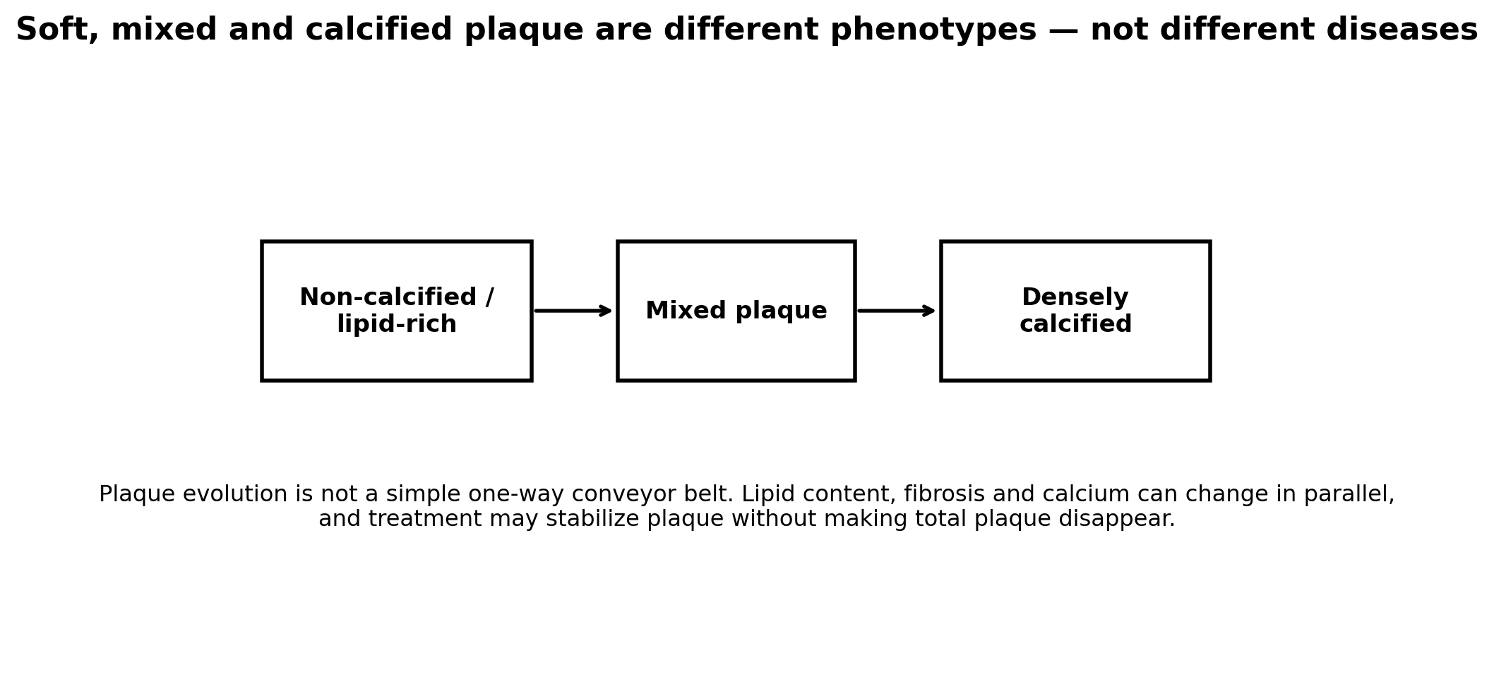
Dense macrocalcification is not simply “bad calcium.” Statin therapy can increase plaque calcification and calcium density while reducing lipid-rich plaque and cardiovascular events—a pattern interpreted as stabilization.
A CAC score can be zero even when non-calcified plaque exists, particularly in younger adults or selected high-risk patients. That is why CAC and CCTA answer different questions.
The best prevention target is not “convert every plaque to calcium.” It is to reduce the number of atherogenic particles, control inflammation and blood pressure, and lower total future event risk.
1. What "soft plaque" means
In everyday language, “soft plaque” usually refers to non-calcified plaque seen on CCTA. It may contain lipid-rich, fibrous or fibrofatty tissue. CCTA attenuation values overlap, so CT cannot reproduce histology perfectly.
Low-attenuation plaque—commonly defined as focal plaque with attenuation below about 30 HU—is more specifically associated with lipid-rich high-risk morphology.
2. What mixed plaque means
Mixed plaque contains both calcified and non-calcified components. It often reflects an intermediate stage of plaque evolution and can still contain biologically active lipid-rich tissue.
The amount and distribution of each component matter more than the word “mixed” alone.
3. What dense calcified plaque means
Densely calcified plaque generally reflects chronic, fibrotic atherosclerosis. It contributes strongly to the Agatston CAC score and is associated with total plaque burden.
At the lesion level, dense calcium is often less rupture-prone than a large lipid-rich necrotic core with a thin fibrous cap. At the patient level, however, a high calcium burden still signals a large amount of coronary atherosclerosis and high future risk.
4. High-risk plaque features on CCTA
The most consistently recognized high-risk CCTA features are low attenuation, positive remodeling, spotty calcification and the napkin-ring sign. CAD-RADS 2.0 requires at least two of these features for the HRP modifier.
These features add prognostic information beyond stenosis, but they are not a reason to stent a non-obstructive lesion simply because it “looks vulnerable.” Management is primarily systemic prevention unless symptoms, ischemia or anatomy indicate otherwise.
5. Why CAC can be zero despite soft plaque
Calcium develops later than some non-calcified plaque. A young adult may have atherosclerosis before mineralization is detectable. Studies of CCTA show that a minority of people with CAC 0 have non-calcified plaque, although event rates remain low overall.
This is why CAC is excellent for risk reclassification but is not an absolute rule-out test for all coronary plaque.
6. What statins do to plaque composition
Serial IVUS, OCT and CCTA studies show that intensive LDL lowering can reduce lipid-rich or low-attenuation plaque, increase fibrous-cap thickness and increase dense calcification. These changes can coexist with only modest reductions in total plaque volume.
Therefore, a rising CAC score after statin initiation should not automatically be interpreted as plaque worsening.
| Plaque phenotype | What it suggests | What it does NOT prove |
|---|---|---|
| Non-calcified | Earlier / lipid-rich or fibrous plaque; invisible to CAC | That rupture is imminent |
| Low-attenuation | Higher-risk lipid-rich morphology | That the lesion needs PCI |
| Mixed | Combination of calcium and non-calcified tissue | A precise “age” of plaque |
| Dense calcified | Chronic plaque burden; often more stable locally | That the patient is low risk |
| CAC 0 | No detectable calcified coronary plaque | No non-calcified plaque exists |
7. FAQ
Is soft plaque more dangerous than calcified plaque?
Some soft-plaque phenotypes are more rupture-prone, but patient risk depends on total plaque burden and systemic risk, not a single lesion label.
Can soft plaque become calcified?
Yes. Plaques often become more fibrotic and calcified over time, and statin therapy can accelerate dense calcification as part of stabilization.
Can CCTA measure soft plaque accurately?
Modern quantitative CCTA can estimate non-calcified and low-attenuation plaque, but measurements depend on scanner quality and analysis method.
Should high-risk plaque be stented?
Not automatically. Non-obstructive high-risk plaque is usually treated with intensive prevention rather than prophylactic PCI.
References
1. Cury RC, Leipsic J, Abbara S, et al. CAD-RADS 2.0 - 2022 Coronary Artery Disease-Reporting and Data System. JACC Cardiovasc Imaging. 2022;15(11):1974-2001. doi:10.1016/j.jcmg.2022.07.002.
2. Ferencik M, Mayrhofer T, Bittner DO, et al. Use of High-Risk Coronary Atherosclerotic Plaque Detection for Risk Stratification of Patients With Stable Chest Pain: A Secondary Analysis of the PROMISE Randomized Clinical Trial. JAMA Cardiol. 2018;3(2):144-152. doi:10.1001/jamacardio.2017.4973.
3. Williams MC, Moss AJ, Dweck M, et al. Coronary Artery Plaque Characteristics Associated With Adverse Outcomes in the SCOT-HEART Study. J Am Coll Cardiol. 2019;73(3):291-301. doi:10.1016/j.jacc.2018.10.066.
4. Motoyama S, Ito H, Sarai M, et al. Plaque Characterization by Coronary Computed Tomography Angiography and the Likelihood of Acute Coronary Events in Mid-Term Follow-Up. J Am Coll Cardiol. 2015;66(4):337-346. doi:10.1016/j.jacc.2015.05.069.
5. Otsuka K, Fukuda S, Tanaka A, et al. Napkin-ring sign on coronary CT angiography for the prediction of acute coronary syndrome. JACC Cardiovasc Imaging. 2013;6(4):448-457. doi:10.1016/j.jcmg.2012.09.016.
6. Puri R, Nicholls SJ, Shao M, et al. Impact of Statins on Serial Coronary Calcification During Atheroma Progression and Regression. J Am Coll Cardiol. 2015;65(13):1273-1282. doi:10.1016/j.jacc.2015.01.036.
7. Lee SE, Chang HJ, Sung JM, et al. Effects of Statins on Coronary Atherosclerotic Plaques: The PARADIGM Study. JACC Cardiovasc Imaging. 2018;11(10):1475-1484. doi:10.1016/j.jcmg.2018.04.015.
8. Vergallo R, Park SJ, Stone GW, et al. Vulnerable or High-Risk Plaque: A JACC: Cardiovascular Imaging Position Statement. JACC Cardiovasc Imaging. 2025;18(6):709-740. doi:10.1016/j.jcmg.2024.12.004.