Publication metadata
| Recommended SEO title | Can Atherosclerotic Plaque Really Regress? Landmark Trials Explained |
|---|---|
| Suggested slug | /can-atherosclerotic-plaque-regress/ |
| Meta description | Can coronary plaque really shrink? A rigorous guide to ASTEROID, SATURN, GLAGOV, PACMAN-AMI, HUYGENS, EVAPORATE, CAC and what plaque stabilization really means. |
| Primary keyword | atherosclerotic plaque regression |
| Search intent | Educational / medical evidence / preventive cardiology |
Executive summary
Yes — coronary atherosclerotic plaque can regress. But the word regression is easy to misunderstand. In the major imaging trials, regression usually meant a modest reduction in plaque volume measured within a specific arterial segment. It did not mean that coronary artery disease vanished, calcium returned to zero, or a previously diseased artery became biologically identical to a never-diseased artery.
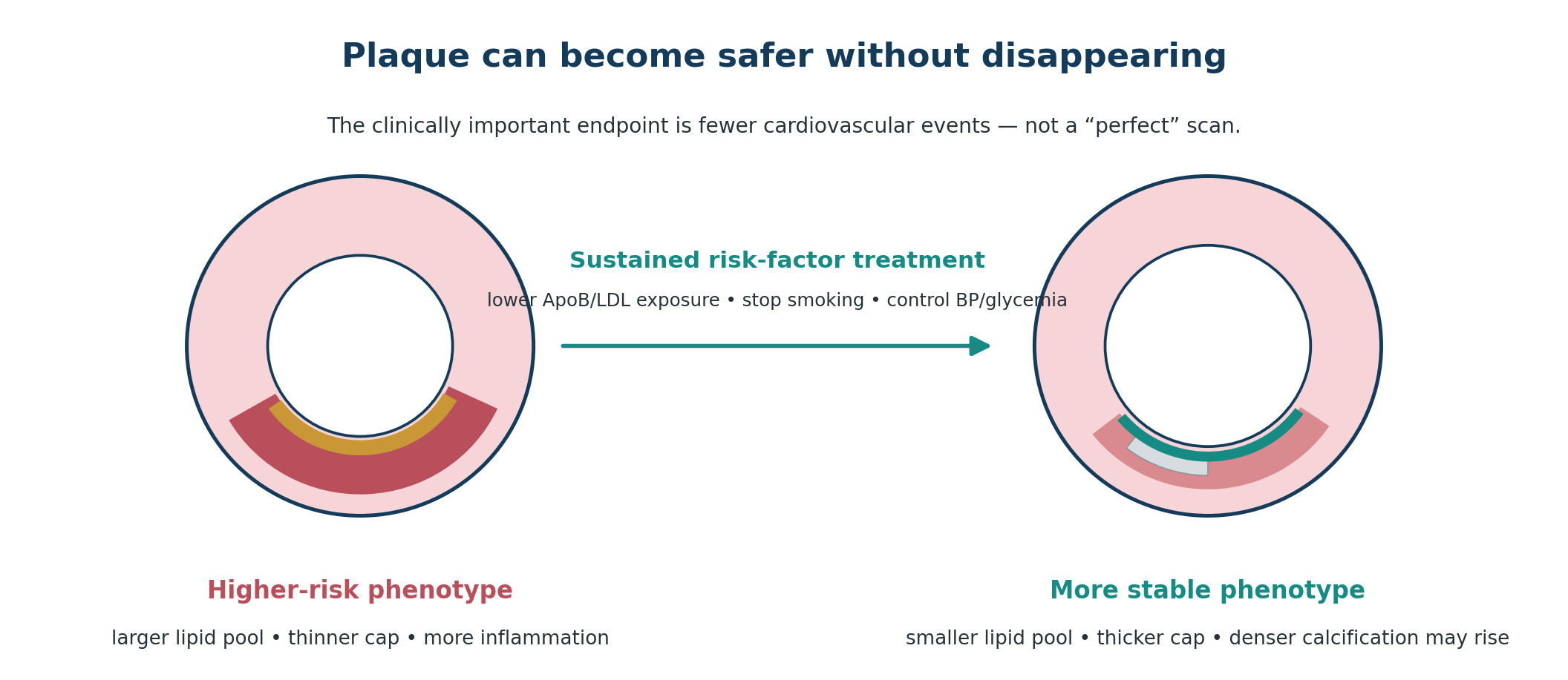
The more important insight from two decades of intravascular ultrasound (IVUS), optical coherence tomography (OCT) and coronary CT angiography (CCTA) is that treatment can change plaque phenotype as well as plaque size. Lipid-rich tissue can contract, fibrous caps can thicken, inflammatory features can diminish, and calcification can become denser. A plaque can therefore become more stable even when its total volume changes only slightly — and even when coronary calcium increases.
The landmark sequence is remarkably coherent. REVERSAL showed that intensive statin therapy could halt progression more effectively than moderate therapy. ASTEROID demonstrated average IVUS-measured regression with rosuvastatin 40 mg. SATURN confirmed regression with two different high-intensity statin regimens. PRECISE-IVUS showed greater regression when ezetimibe was added to atorvastatin in a selected PCI population. GLAGOV moved LDL-C into the 30s with evolocumab and produced additional regression. PACMAN-AMI and HUYGENS then showed that PCSK9 inhibition could alter not only plaque volume but lipid burden and fibrous-cap structure after myocardial infarction. [5-12]
These imaging results align with large outcome trials showing that therapies that lower atherogenic lipoprotein exposure — statins, ezetimibe and PCSK9 inhibition — reduce myocardial infarction and stroke. Imaging explains part of the biology; outcome trials establish clinical benefit. [13-17]
The practical goal is therefore not to obtain a prettier scan. It is to reduce lifetime exposure to ApoB-containing particles, control blood pressure and metabolic risk, avoid smoking, use evidence-based therapies when indicated, and convert the arterial environment from one that promotes lipid-rich vulnerable plaque to one that is less likely to rupture.
Plaque regression is real, but plaque stabilization is the bigger story. A small reduction in plaque volume plus a thicker fibrous cap, less lipid and fewer inflammatory features may be more clinically meaningful than a dramatic change in lumen size.
At a glance: what the landmark trials changed
| Trial | Imaging | Intervention | Main signal | Why it mattered |
|---|---|---|---|---|
| REVERSAL (2004) | IVUS | Atorvastatin 80 vs pravastatin 40 | Intensive therapy halted progression more effectively | Established that intensity of LDL lowering changes plaque biology |
| ASTEROID (2006) | IVUS | Rosuvastatin 40 | Mean PAV −0.98 percentage points | Clear early demonstration of average coronary plaque regression |
| SATURN (2011) | IVUS | Rosuvastatin 40 vs atorvastatin 80 | Both groups regressed | Regression was not unique to one statin |
| PRECISE-IVUS (2015) | IVUS | Atorvastatin + ezetimibe vs atorvastatin | Greater PAV regression with combination | Supported deeper LDL lowering using a non-statin add-on |
| GLAGOV (2016) | IVUS | Evolocumab + statin vs placebo + statin | PAV −0.95% vs +0.05% | Very low LDL produced additional regression |
| PARADIGM (2018) | CCTA | Observed statin use | Slower noncalcified progression; more calcification | Explained why calcium can rise as plaque stabilizes |
| EVAPORATE (2020) | CCTA | Icosapent ethyl 4 g/day vs placebo | Lower low-attenuation plaque in a small trial | Suggested favorable plaque-composition effects of purified EPA |
| PACMAN-AMI (2022) | IVUS/NIRS/OCT | Alirocumab + statin vs placebo + statin | More regression, less lipid, thicker caps | Showed simultaneous improvement in plaque quantity and quality |
| HUYGENS (2022) | OCT/IVUS | Evolocumab + statin vs placebo + statin | Greater fibrous-cap thickening; less lipid arc | Strengthened the plaque-stabilization model |
1. What does "plaque regression" actually mean?
Atherosclerosis develops within the arterial wall, not simply inside the lumen. ApoB-containing lipoproteins enter and become retained in the intima; inflammatory cells accumulate; extracellular lipid and necrotic material expand; smooth-muscle cells and collagen form a fibrous cap; and calcium is deposited as disease evolves. The resulting plaque can enlarge outward — positive remodeling — for years before angiography shows major luminal narrowing. [1-3]
That anatomy is why a person can have biologically important atherosclerosis with only mild stenosis, and why successful treatment may not produce a dramatic increase in lumen diameter. When investigators talk about regression, they usually quantify the amount of tissue between the lumen and the outer vessel boundary over a defined segment. A reduction of around one percentage point in percent atheroma volume (PAV) is scientifically meaningful in a trial even though it sounds small in everyday language.
Regression also differs from stabilization. A plaque that remains similar in overall size can become less dangerous if its lipid pool contracts, inflammatory activity falls and the fibrous cap becomes thicker. Conversely, a plaque could theoretically shrink without becoming fully benign. Modern imaging studies therefore increasingly measure both burden and composition.
Myth: “Regression” means the plaque dissolves. Fact: In trials, regression usually means a modest reduction in measured atheroma volume. The artery remains atherosclerotic, and risk reduction depends on the whole vascular system, not one plaque.
2. How researchers measure plaque
No single imaging method sees every feature of atherosclerosis. The apparent amount of “regression” depends partly on what is being measured.
| Method | What it sees best | Strength | Key limitation for follow-up |
|---|---|---|---|
| IVUS | Plaque burden and vessel dimensions | Validated serial measurement of PAV/TAV | Invasive; limited tissue characterization compared with OCT |
| OCT | Fibrous cap, lipid arc, macrophage-like signals | Very high axial resolution | Invasive; evaluates selected coronary segments |
| NIRS | Lipid-core burden | Quantifies lipid-rich tissue | Invasive; often combined with IVUS |
| CCTA | Whole-tree calcified and noncalcified plaque, stenosis | Noninvasive anatomic overview | Scanner/protocol/software dependence; radiation/contrast |
| CAC | Calcified plaque burden only | Excellent risk stratification in selected asymptomatic adults | Cannot see noncalcified plaque or measure treatment response reliably |
IVUS created the modern plaque-regression literature because it can trace the lumen and external elastic membrane along the same coronary segment at baseline and follow-up. PAV expresses plaque volume as a proportion of vessel volume. TAV is an absolute volume measure. OCT later added a different question: is the fibrous cap becoming thicker and the lipid-rich plaque less vulnerable? CCTA extended the concept noninvasively to the wider coronary tree. [5-12]
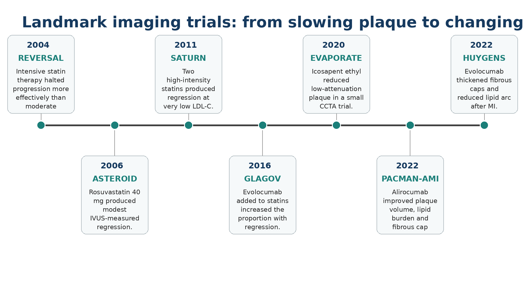
Figure 1. Selected imaging trials that shifted the field from “slowing plaque” to regression and stabilization. Original editorial figure.
3. The landmark trials --- what each one actually proved
3.1 REVERSAL: first, stop progression
REVERSAL randomized patients with coronary disease to intensive atorvastatin 80 mg or moderate pravastatin 40 mg and followed plaque with IVUS for 18 months. LDL-C fell to about 79 mg/dL with atorvastatin and 110 mg/dL with pravastatin. Total atheroma volume changed by approximately −0.4% with intensive treatment versus +2.7% with moderate treatment. The study’s name was aspirational: the strongest message was that intensive therapy could arrest progression far more effectively than moderate treatment. [5]
REVERSAL also lowered hs-CRP more with intensive atorvastatin, helping to establish the modern idea that aggressive lipid lowering changes both atherogenic exposure and the inflammatory environment. But the trial was not designed to show that a specific percentage of plaque reduction translates directly into a specific reduction in myocardial infarction.
3.2 ASTEROID: average plaque volume moved backward
ASTEROID was the study that made the phrase plaque regression mainstream. Five hundred seven patients received rosuvastatin 40 mg daily; 349 had evaluable serial IVUS after 24 months. Mean LDL-C fell from 130.4 to 60.8 mg/dL, while HDL-C rose. Mean PAV declined by 0.98 percentage points and normalized total atheroma volume fell by a median 6.8%. [6]
This was compelling mechanistic evidence, but ASTEROID had no placebo or active-control group. It therefore demonstrated that regression occurred during very intensive statin therapy, not that the observed imaging change by itself caused fewer events. The correct lesson is stronger than the marketing version: coronary plaque is biologically dynamic, and sustained intensive LDL lowering can move average atheroma burden in the favorable direction.
3.3 SATURN: regression was reproducible across high-intensity statins
SATURN directly compared rosuvastatin 40 mg with atorvastatin 80 mg in more than 1,000 patients with coronary disease. Achieved LDL-C was lower with rosuvastatin (about 62.6 mg/dL versus 70.2 mg/dL), but both groups showed significant IVUS regression. Mean PAV fell by roughly 1% in both groups; regression was observed in 68.5% of rosuvastatin-treated participants and 63.2% of atorvastatin-treated participants. [7]
The importance of SATURN is conceptual. Plaque regression was not a unique property of one molecule. It was reproducible with two maximally intensive statin strategies. That supports a broader exposure model: the lower the burden of circulating atherogenic particles over time, the more favorable the arterial response tends to be.
3.4 PRECISE-IVUS: deeper LDL lowering with ezetimibe
PRECISE-IVUS randomized 202 Japanese patients who had undergone PCI to atorvastatin alone or atorvastatin plus ezetimibe. LDL-C averaged 63.2 mg/dL with combination therapy versus 73.3 mg/dL with atorvastatin alone. PAV fell by 1.4% with the combination versus 0.3% with atorvastatin alone, and 78% versus 58% met the trial definition of plaque regression. [8]
This was a relatively small imaging trial, but its direction matched the much larger IMPROVE-IT outcome trial, in which adding ezetimibe to simvastatin after acute coronary syndrome lowered LDL-C further and modestly reduced cardiovascular events. Imaging and outcomes therefore pointed in the same direction: additional reduction of ApoB-containing lipoprotein exposure can add benefit. [13]
3.5 GLAGOV: what happens when LDL reaches the 30s?
GLAGOV randomized 968 statin-treated patients with angiographic coronary disease to evolocumab or placebo for 76 weeks. Mean time-weighted LDL-C was 36.6 mg/dL with evolocumab and 93.0 mg/dL with placebo. PAV decreased by 0.95 percentage points with evolocumab but increased by 0.05 with placebo. Regression by PAV occurred in 64.3% versus 47.3% of patients; by normalized TAV, 61.5% versus 48.9%. [9]
This is where social-media shorthand can become misleading. GLAGOV did not show that evolocumab erased 40% of everyone’s plaque. It showed modest average IVUS regression and a substantially larger proportion of patients meeting a predefined regression criterion. The clinical significance of PCSK9 inhibition was then established by outcomes trials such as FOURIER and ODYSSEY OUTCOMES, which demonstrated fewer ischemic events with profound LDL lowering. [14,15]
3.6 PACMAN-AMI: shrinking plaque and changing its structure
PACMAN-AMI moved beyond a single measurement. Three hundred patients with acute myocardial infarction received high-intensity statin therapy and were randomized to alirocumab or placebo. After 52 weeks, alirocumab produced greater PAV regression in non-infarct-related arteries (−2.13% versus −0.92%), a larger fall in lipid-core burden by near-infrared spectroscopy, and a greater increase in minimal fibrous-cap thickness by OCT. [10]
This is one of the clearest demonstrations that “plaque treatment” is multidimensional. The plaque became smaller, less lipid-rich and more heavily protected by fibrous tissue. That biological package is much closer to what clinicians mean by stabilization than a simple change in stenosis percentage.
3.7 HUYGENS: fibrous caps can thicken rapidly
HUYGENS enrolled statin-treated patients after non–ST-segment elevation myocardial infarction and randomized them to evolocumab or placebo for 52 weeks. LDL-C fell to approximately 28 mg/dL with evolocumab versus 87 mg/dL with placebo. Evolocumab produced a greater increase in minimum fibrous-cap thickness (+42.7 versus +21.5 μm) and a larger reduction in lipid arc. [11]
The study reinforced an important principle already suggested by the smaller EASY-FIT trial: a lipid-rich plaque can become mechanically more robust within months when atherogenic exposure is driven down. [18] Plaque biology can therefore improve faster than the casual observer might expect from the slow natural history of calcified disease.
3.8 EVAPORATE: a CCTA signal for purified EPA
EVAPORATE randomized only 80 statin-treated patients with coronary disease and fasting triglycerides of 135–499 mg/dL to icosapent ethyl 4 g/day or placebo. Over 18 months, low-attenuation plaque volume fell 17% in the icosapent ethyl group while increasing 109% in the placebo group; other noncalcified plaque components also moved favorably. [12]
The result is intriguing because low-attenuation plaque is a CCTA marker associated with future myocardial infarction risk. But EVAPORATE was small and was not an outcomes trial. The reason icosapent ethyl has a role in selected high-risk patients is primarily the REDUCE-IT outcomes evidence, not the promise that every patient taking fish oil will shrink plaque. REDUCE-IT tested prescription icosapent ethyl, not generic mixed EPA/DHA supplements. [16]
4. Why coronary calcium can increase while plaque becomes safer
This is one of the most counterintuitive findings in preventive cardiology. Statins reduce cardiovascular events and can regress atheroma, yet serial imaging often shows more plaque calcification. That is not a contradiction.
A pooled analysis of eight IVUS trials found that high-intensity statin therapy was associated with PAV regression but also with increased indices of coronary calcification. [19] PARADIGM, a large serial-CCTA registry, similarly found slower overall and noncalcified plaque progression among statin-treated patients but faster progression of calcified plaque and fewer newly developing high-risk plaque features. [20]
One plausible interpretation is that lipid-rich, biologically active plaque is being transformed toward a denser, more organized calcified phenotype. Dense calcium is not harmless — it proves atherosclerosis exists — but the transition can be part of stabilization. The same patient can therefore have a higher Agatston calcium score and a lower risk than they would have had without treatment.
This is why serial CAC scanning is a poor way to judge whether a statin, ezetimibe or PCSK9 inhibitor is “working.” CAC is excellent for risk stratification in appropriately selected untreated or asymptomatic patients, but change in CAC after treatment has not been validated as a therapeutic target.
Do not interpret a rising CAC score on therapy as proof that plaque is getting more dangerous. Treatment can increase calcified plaque density while reducing lipid-rich plaque and clinical events.
5. Does imaging regression translate into fewer heart attacks?
This is the question that separates mechanistic enthusiasm from evidence-based medicine. Imaging trials are generally small, select patients who can undergo repeated invasive imaging, and are not powered for mortality or myocardial infarction. A beautiful IVUS or OCT result is not sufficient to recommend a drug.
The strongest case arises when an imaging signal and an independent cardiovascular-outcomes program agree. Statins produce regression and reduce events. Ezetimibe can enhance regression in selected imaging cohorts and reduced events in IMPROVE-IT. PCSK9 monoclonal antibodies produced additional regression and plaque stabilization in GLAGOV, PACMAN-AMI and HUYGENS, while FOURIER and ODYSSEY OUTCOMES independently demonstrated reductions in major ischemic events. [9-15]
A 2023 systematic review and meta-regression of plaque-regression studies found that greater atherosclerotic regression was associated at the study level with lower adverse cardiovascular-event rates. That supports biological plausibility, but it still does not make plaque change a validated surrogate endpoint for every therapy. [21]
The hierarchy therefore remains: randomized cardiovascular outcomes first; validated risk factors and biomarkers second; imaging as a powerful mechanistic and risk-stratification tool.
6. What actually drives favorable plaque biology?
The trials do not support a single “plaque-dissolving” intervention. They support sustained modification of the exposures that build and destabilize plaque.
Lower ApoB-containing lipoprotein exposure: This is the most reproducible lever. Statins remain foundational; ezetimibe, PCSK9 monoclonal antibodies, bempedoic acid and other approved agents can be combined according to risk, tolerance and guideline targets. The 2026 ACC/AHA multisociety dyslipidemia guideline again emphasizes explicit LDL-C goals, including <55 mg/dL for very-high-risk secondary prevention. [22]
Stop smoking: Smoking accelerates endothelial dysfunction, thrombosis and plaque instability. No imaging supplement compensates for continued tobacco exposure.
Control blood pressure and diabetes: Mechanical stress, hyperglycemia and insulin resistance amplify vascular injury and absolute event risk. Atherosclerosis treatment is not only lipid treatment.
Exercise and cardiorespiratory fitness: Regular physical activity improves blood pressure, insulin sensitivity, endothelial function and overall cardiovascular risk. Small imaging studies suggesting step-count thresholds are hypothesis-generating; they do not prove that a particular number such as 7,000 steps “reduces plaque by 12.5%.”
Use icosapent ethyl only where evidence fits: In high- or very-high-risk statin-treated patients with persistently elevated triglycerides, high-dose prescription icosapent ethyl has randomized outcomes evidence and is supported in current European guidance. This is different from recommending generic fish-oil supplements to everyone. [16,23]
Treat inflammatory risk selectively: Inflammation is integral to atherosclerosis, but anti-inflammatory drugs are not substitutes for lipid lowering. Colchicine and canakinumab trials show that particular pathways can reduce events in selected secondary-prevention settings; the choice depends on indication, contraindications and guideline context.
7. What has not been proven to regress coronary plaque
The popularity of plaque imaging has created a parallel market for products advertised as “artery cleaners.” The evidentiary standard should be much higher.
| Claim | Current evidence | Editorial interpretation |
|---|---|---|
| Vitamin K2 “removes coronary calcium” | Randomized CAC data are emerging, but no established evidence that K2 removes plaque or prevents ASCVD events | Interesting research question; not a replacement for proven LDL/ApoB and BP treatment |
| Nattokinase “dissolves plaque” | Limited small human studies; no robust major cardiovascular outcome evidence | Insufficient for routine plaque-regression therapy; bleeding interactions matter |
| Serrapeptase “breaks down arterial fibrin” | No persuasive randomized coronary plaque or outcome evidence | Do not present as evidence-based atherosclerosis treatment |
| IV phosphatidylcholine / Plaquex “cleans arteries” | No modern large randomized evidence establishing coronary plaque regression or event reduction | Experimental/integrative claim, not standard preventive cardiology |
| Generic fish oil equals icosapent ethyl | Formulations and trial results differ | Do not extrapolate REDUCE-IT/EVAPORATE to all over-the-counter omega-3 products |
8. Should you repeat CAC or CCTA to see whether plaque regressed?
Usually, not simply for reassurance. A treatment-response test is useful only if the result is reliable, interpretable and changes management. Serial CAC fails the second criterion because calcification can increase during successful statin therapy. A higher score does not tell you whether lipid-rich plaque has fallen or whether the plaque has stabilized. [19,20]
Serial CCTA is more informative because it can quantify noncalcified plaque and high-risk features, but routine repeated CCTA in asymptomatic people solely to document regression is not established as an outcomes-improving strategy. Measurements can vary with scanner, heart rate, contrast timing, reconstruction method and software. Radiation and iodinated contrast are additional considerations.
In research, serial CCTA is extraordinarily useful. In clinical care, a repeat scan should answer a specific question: new or changing symptoms, uncertainty about anatomy, or a management decision that cannot be resolved more simply. The 2021 chest-pain guideline supports CCTA primarily as a diagnostic test in appropriate symptomatic risk groups, not as a universal annual plaque tracker. [24]
For most patients, the most actionable follow-up remains simpler: adherence, LDL-C/non-HDL-C/ApoB, blood pressure, glycemia, smoking status, symptoms, exercise capacity and medication tolerance.
9. A practical evidence-based framework
Establish whether atherosclerosis is present and how the person’s overall risk is classified. A CAC score, CCTA, prior myocardial infarction, stroke or revascularization do not carry the same implications.
Quantify modifiable atherogenic exposure with LDL-C, non-HDL-C and — particularly when discordance is possible — ApoB. Elevated Lp(a) is an additional inherited risk amplifier, not a reason to ignore ApoB lowering.
Set lipid goals according to global risk and current guidelines. In very-high-risk secondary prevention, both contemporary U.S. and European guidance support very low LDL-C targets. [22,23]
Use therapies with cardiovascular-outcomes evidence first. Imaging evidence is a useful supporting layer, not a substitute for proven event reduction.
Control the rest of the arterial environment: smoking, blood pressure, diabetes, visceral adiposity, physical inactivity and diet quality.
Do not use rising CAC as a reason to stop statin therapy. If treatment is well tolerated and appropriately indicated, the calcium change is not a valid measure of failure.
Repeat anatomic imaging only when it is likely to answer a clinically meaningful question.
Success is not “zero plaque.” It is lower event probability: fewer ApoB particles reaching the artery wall, less lipid-rich and inflammatory plaque, thicker fibrous caps, controlled blood pressure and metabolic risk, and long-term adherence to therapies with proven benefit.
10. Frequently asked questions
Can plaque disappear completely?
Small plaques may become difficult to detect, but established coronary atherosclerosis should not be thought of as a reversible skin lesion. Trials demonstrate average regression and stabilization, not restoration of every diseased artery to a pristine state.
How fast can plaque change?
Some structural changes can be detected within 6–12 months in research studies, particularly fibrous-cap thickening after intensive lipid lowering. Larger changes in overall atheroma burden usually require longer exposure.
If my stenosis is only 10%, does plaque matter?
Yes. Stenosis describes lumen narrowing, not total plaque biology. Positive remodeling can preserve the lumen despite substantial plaque burden, and many acute events arise from lesions that were not severely obstructive beforehand.
Is calcified plaque safer than soft plaque?
Dense calcification is generally a more stable phenotype than lipid-rich low-attenuation plaque, but calcium is still evidence of atherosclerotic disease. “More stable” does not mean “normal.”
Can lowering LDL too much be harmful?
Large PCSK9 outcome trials have achieved very low LDL-C levels without a clear signal of major neurocognitive or hemorrhagic-stroke harm over trial follow-up. Targets should still be individualized, but modern evidence supports intensive lowering in high-risk patients. [14,15,22]
What about high Lp(a)?
Lp(a) is an ApoB-containing, genetically determined risk factor. Until dedicated outcome-proven Lp(a)-lowering therapy is established, the most actionable approach is aggressive control of modifiable ApoB/LDL exposure and every other risk factor.
Do I need a PCSK9 inhibitor to regress plaque?
No. Statin trials demonstrated regression before PCSK9 inhibitors existed. PCSK9 inhibitors add substantial LDL lowering and can produce additional regression in appropriately selected patients, but treatment choice depends on risk, achieved LDL-C/ApoB, tolerance, access and guideline indications.
Is plaque regression the same as reversing heart disease?
No. It is one measurable component of modifying a chronic systemic disease. The clinically meaningful definition of “reversal” would be sustained reduction in cardiovascular events, not merely a change on a scan.
Key take-home messages
Coronary plaque regression is real, but the average changes in controlled imaging studies are usually modest.
Plaque stabilization — less lipid, thicker fibrous caps and fewer high-risk features — may be as important as a reduction in total plaque volume.
REVERSAL, ASTEROID and SATURN established the statin regression story; GLAGOV, PACMAN-AMI and HUYGENS showed that deeper LDL lowering with PCSK9 inhibition can move plaque further.
CAC can increase during successful lipid-lowering therapy. Serial CAC is not a reliable treatment-response test.
CCTA can show noncalcified and low-attenuation plaque, but routine serial CCTA solely to document regression is not established standard care.
Outcome trials matter more than imaging trials when deciding whether a therapy should be used.
The most evidence-based way to “treat plaque” is sustained control of ApoB/LDL exposure plus smoking, blood pressure, diabetes, physical activity and other global risk factors.
Be skeptical of supplements or infusions marketed as plaque dissolvers when they lack modern randomized cardiovascular outcome evidence.
References
1. Libby P. The changing landscape of atherosclerosis. Nature. 2021;592:524-533. doi:10.1038/s41586-021-03392-8.
2. Ference BA, Ginsberg HN, Graham I, et al. Low-density lipoproteins cause atherosclerotic cardiovascular disease. Eur Heart J. 2017;38:2459-2472. doi:10.1093/eurheartj/ehx144.
3. Stone GW, Maehara A, Lansky AJ, et al. A prospective natural-history study of coronary atherosclerosis. N Engl J Med. 2011;364:226-235. doi:10.1056/NEJMoa1002358.
4. Puri R, et al. Coronary atherosclerotic plaque regression: JACC state-of-the-art review. J Am Coll Cardiol. 2022;79:66-82. doi:10.1016/j.jacc.2021.10.035.
5. Nissen SE, Tuzcu EM, Schoenhagen P, et al. Effect of intensive compared with moderate lipid-lowering therapy on progression of coronary atherosclerosis: a randomized controlled trial. JAMA. 2004;291:1071-1080. doi:10.1001/jama.291.9.1071.
6. Nissen SE, Nicholls SJ, Sipahi I, et al. Effect of very high-intensity statin therapy on regression of coronary atherosclerosis: the ASTEROID trial. JAMA. 2006;295:1556-1565. doi:10.1001/jama.295.13.jpc60002.
7. Nicholls SJ, Ballantyne CM, Barter PJ, et al. Effect of two intensive statin regimens on progression of coronary disease. N Engl J Med. 2011;365:2078-2087. doi:10.1056/NEJMoa1110874.
8. Tsujita K, Sugiyama S, Sumida H, et al. Impact of dual lipid-lowering strategy with ezetimibe and atorvastatin on coronary plaque regression: PRECISE-IVUS. J Am Coll Cardiol. 2015;66:495-507. doi:10.1016/j.jacc.2015.05.065.
9. Nicholls SJ, Puri R, Anderson T, et al. Effect of evolocumab on progression of coronary disease in statin-treated patients: the GLAGOV randomized clinical trial. JAMA. 2016;316:2373-2384. doi:10.1001/jama.2016.16951.
10. Räber L, Ueki Y, Otsuka T, et al. Effect of alirocumab added to high-intensity statin therapy on coronary atherosclerosis in patients with acute myocardial infarction: PACMAN-AMI. JAMA. 2022;327:1771-1781. doi:10.1001/jama.2022.5218.
11. Nicholls SJ, Kataoka Y, Nissen SE, et al. Effect of evolocumab on coronary plaque phenotype and burden in statin-treated patients following myocardial infarction. JACC Cardiovasc Imaging. 2022;15:1308-1321. doi:10.1016/j.jcmg.2022.03.002.
12. Budoff MJ, Bhatt DL, Kinninger A, et al. Effect of icosapent ethyl on progression of coronary atherosclerosis in patients with elevated triglycerides on statin therapy: final results of the EVAPORATE trial. Eur Heart J. 2020;41:3925-3932. doi:10.1093/eurheartj/ehaa652.
13. Cannon CP, Blazing MA, Giugliano RP, et al. Ezetimibe added to statin therapy after acute coronary syndromes. N Engl J Med. 2015;372:2387-2397. doi:10.1056/NEJMoa1410489.
14. Sabatine MS, Giugliano RP, Keech AC, et al. Evolocumab and clinical outcomes in patients with cardiovascular disease. N Engl J Med. 2017;376:1713-1722. doi:10.1056/NEJMoa1615664.
15. Schwartz GG, Steg PG, Szarek M, et al. Alirocumab and cardiovascular outcomes after acute coronary syndrome. N Engl J Med. 2018;379:2097-2107. doi:10.1056/NEJMoa1801174.
16. Bhatt DL, Steg PG, Miller M, et al. Cardiovascular risk reduction with icosapent ethyl for hypertriglyceridemia. N Engl J Med. 2019;380:11-22. doi:10.1056/NEJMoa1812792.
17. Cholesterol Treatment Trialists’ Collaboration. Efficacy and safety of more intensive lowering of LDL cholesterol: meta-analysis of data from 170,000 participants in 26 randomized trials. Lancet. 2010;376:1670-1681. doi:10.1016/S0140-6736(10)61350-5.
18. Komukai K, Kubo T, Kitabata H, et al. Effect of atorvastatin therapy on fibrous cap thickness in coronary atherosclerotic plaque as assessed by optical coherence tomography: the EASY-FIT study. J Am Coll Cardiol. 2014;64:2207-2217. doi:10.1016/j.jacc.2014.08.045.
19. Puri R, Nicholls SJ, Shao M, et al. Impact of statins on serial coronary calcification during atheroma progression and regression. J Am Coll Cardiol. 2015;65:1273-1282. doi:10.1016/j.jacc.2015.01.036.
20. Lee SE, Chang HJ, Sung JM, et al. Effects of statins on coronary atherosclerotic plaques: the PARADIGM study. JACC Cardiovasc Imaging. 2018;11:1475-1484. doi:10.1016/j.jcmg.2018.04.015.
21. Mancini GBJ, et al. Atherosclerotic coronary plaque regression and risk of adverse cardiovascular events: a systematic review and updated meta-regression analysis. JAMA Cardiol. 2023;8:937-946. doi:10.1001/jamacardio.2023.2731.
22. Blumenthal RS, Morris PB, Gaudino M, et al. 2026 ACC/AHA/AACVPR/ABC/ACPM/ADA/AGS/APhA/ASPC/NLA/PCNA guideline on the management of dyslipidemia. Circulation. Published online March 13, 2026. doi:10.1161/CIR.0000000000001423.
23. Mach F, Koskinas KC, Roeters van Lennep JE, et al. 2025 focused update of the 2019 ESC/EAS Guidelines for the management of dyslipidaemias. Eur Heart J. 2025. doi:10.1093/eurheartj/ehaf190.
24. Gulati M, Levy PD, Mukherjee D, et al. 2021 AHA/ACC/ASE/CHEST/SAEM/SCCT/SCMR guideline for the evaluation and diagnosis of chest pain. Circulation. 2021;144:e368-e454. doi:10.1161/CIR.0000000000001029.
25. Williams MC, Kwiecinski J, Doris M, et al. Low-attenuation noncalcified plaque on coronary CT angiography predicts myocardial infarction: results from SCOT-HEART. Circulation. 2020;141:1452-1462.