Executive summary
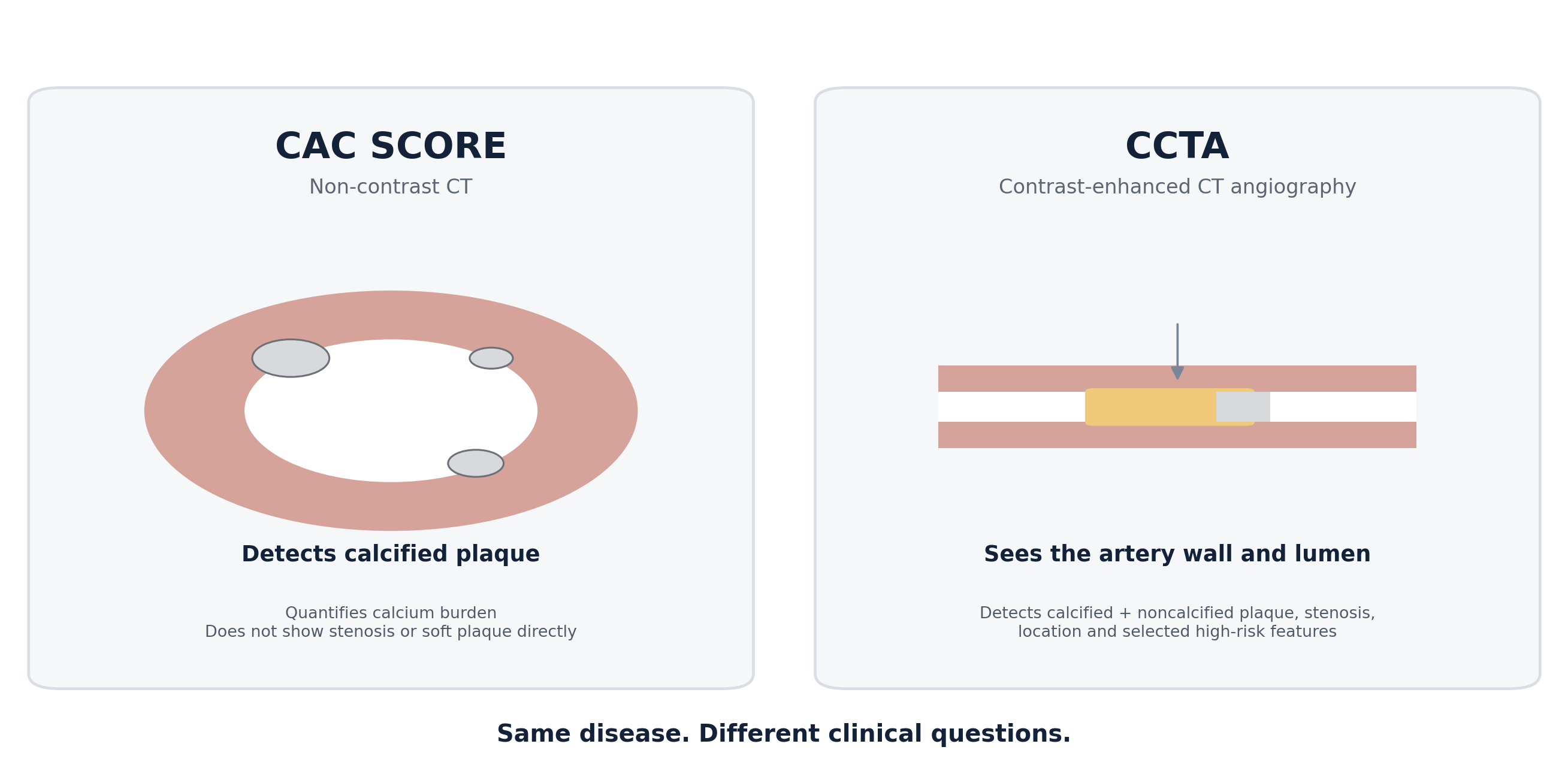
Coronary artery calcium (CAC) scoring and coronary CT angiography (CCTA) are often discussed as if one were simply a more advanced version of the other. They are not. They answer different questions. CAC is a fast, non-contrast measure of calcified coronary plaque burden and is exceptionally useful for refining risk in selected asymptomatic adults. CCTA is a contrast-enhanced anatomical examination that can show the coronary lumen and vessel wall, identify calcified and noncalcified plaque, characterize stenosis, and—in appropriate settings—identify plaque features associated with higher risk. [1–4]
The simplest distinction is this: CAC asks, “How much calcified coronary atherosclerosis is present?” CCTA asks, “What does the coronary artery disease actually look like?” That difference matters because plaque biology evolves over time. Early and lipid-rich plaque may be noncalcified. Later disease often contains mixed or dense calcium. A person can therefore have CAC=0 and still have noncalcified plaque, although in asymptomatic populations the overall prevalence of such plaque is relatively low and obstructive disease is uncommon. [7–9]
CAC and CCTA are complementary, not interchangeable. The best test is the one that answers the clinical question with the least unnecessary testing.
CAC vs CCTA at a glance
| Question | CAC score | CCTA |
|---|---|---|
| Contrast required? | No | Yes — iodinated contrast |
| Main output | Agatston calcium score / percentile | Coronary anatomy, plaque and stenosis |
| Calcified plaque | Yes | Yes |
| Noncalcified / “soft” plaque | No direct visualization | Yes |
| Luminal stenosis | No | Yes |
| High-risk plaque features | No | Selected features can be assessed |
| Best-established role | Risk refinement in selected asymptomatic primary prevention | Diagnosis/risk stratification in suspected chronic coronary syndrome and selected known CAD |
| Typical practical advantage | Fast, inexpensive, low radiation, no contrast | Much richer anatomical information |
| Major limitation | Does not show soft plaque or stenosis | Contrast, more preparation, artifacts; not a routine population-screening test |
1. Start with the question, not the scanner
Cardiovascular imaging is most useful when the clinical question is explicit. An asymptomatic 55-year-old with borderline treatment uncertainty is a very different scenario from a 55-year-old with exertional chest pressure. In the first setting, the question may be whether subclinical atherosclerosis is present strongly enough to change preventive therapy. In the second, the question is whether coronary artery disease is causing symptoms and whether there is obstructive or otherwise important anatomy. Current guidelines reflect this distinction. [1,2,17]
If the problem is risk uncertainty in an otherwise asymptomatic primary-prevention patient, CAC is often the more appropriate first imaging tool.
If the problem is suspected chronic coronary syndrome or stable chest pain requiring anatomical diagnosis, CCTA is often the more informative test in appropriately selected patients.
If the problem is whether a stenosis is causing ischemia, an anatomical test may need to be complemented by functional testing or CT-derived fractional flow reserve (FFR-CT) in selected cases.
More information is not automatically better information. A technically sophisticated test is useful only if the result can change diagnosis, prognosis or management.
2. What a CAC score actually measures
A CAC scan is a non-contrast ECG-gated CT examination that identifies calcium within the coronary arteries. The most widely used result is the Agatston score, which combines the area and density of calcified lesions into a single number. CAC is therefore not a direct measurement of stenosis and not a complete measurement of total plaque volume. It is a marker of calcified atherosclerotic burden. [5,17]
Common CAC categories
| CAC score | Common description | General interpretation |
|---|---|---|
| 0 | No detectable calcium | Powerful negative risk marker; does not guarantee absence of plaque |
| 1–99 | Mild calcified plaque burden | Atherosclerosis is present; age and percentile matter |
| 100–299 | Moderate burden | Clearly abnormal; generally supports more intensive prevention |
| ≥300 | High burden | Higher long-term event risk; often treated as substantial subclinical disease |
| ≥1000 | Very high burden | Very high plaque burden and risk; may alter intensity of prevention and evaluation |
These categories are useful shorthand, not biological boundaries. Age, sex and percentile can materially change the interpretation. A CAC of 80 in a younger adult may represent a very high age/sex percentile, while the same absolute score in an older person may be less unusual. Likewise, CAC burden must be interpreted alongside diabetes, smoking, blood pressure, ApoB/LDL-C, family history, Lp(a) and other risk enhancers.
The strength of CAC comes from prognosis. In MESA, 10-year ASCVD event rates were low in people with CAC=0 and substantially higher in those with CAC >300 across demographic subgroups. [5] CAC=0 has repeatedly proved to be one of the strongest negative risk markers available in primary prevention. [6]
Myth: “A CAC score tells me how blocked my arteries are.”
Fact: CAC quantifies coronary calcium. A high score strongly implies
atherosclerotic burden, but it does not tell you the percentage
narrowing of any particular artery.
3. The power---and limits---of CAC=0
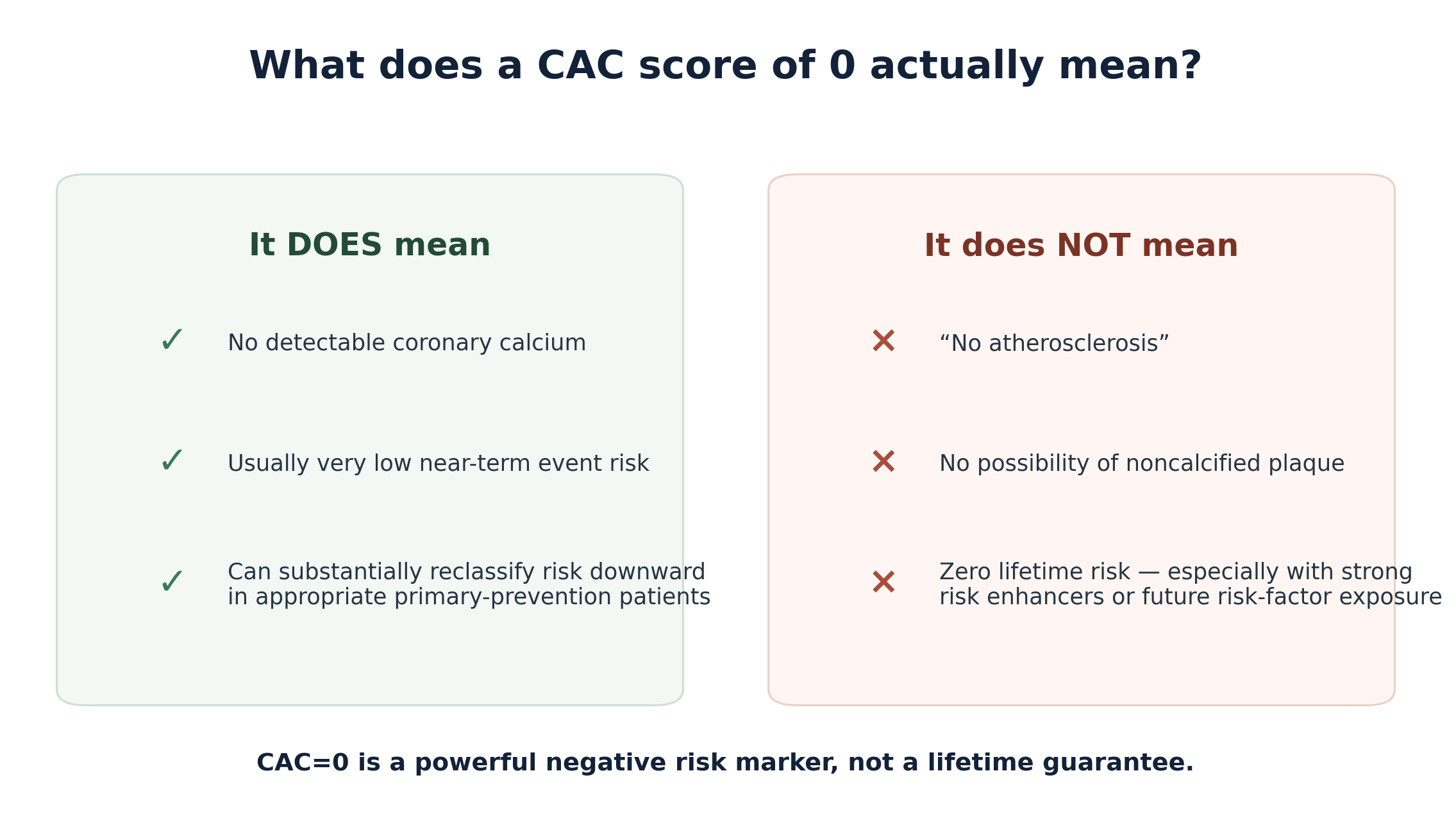
CAC=0 deserves respect because it meaningfully lowers near-term risk in many asymptomatic adults. It is one reason calcium scoring can prevent both overtreatment and undertreatment when treatment decisions are uncertain. But the phrase “power of zero” is sometimes stretched into “zero plaque,” and those statements are not equivalent. [6–10]
Calcification is generally a later component of plaque biology. CCTA studies show that some people with CAC=0 have noncalcified plaque. A 2024 systematic review and meta-analysis of 14 studies involving 37,808 asymptomatic people with CAC=0 estimated a pooled prevalence of any noncalcified plaque of about 10% and obstructive noncalcified plaque of about 1.1%. [7] In symptomatic cohorts the prevalence can be higher, which is why symptoms and pre-test likelihood matter.
The SCOT-HEART imaging analyses reinforce the nuance: zero calcium was associated with a good prognosis, yet did not exclude obstructive disease, nonobstructive plaque or low-attenuation plaque. [9] Thus CAC=0 is best understood as a strong short- to intermediate-term risk modifier rather than a certificate of lifetime immunity.
A CAC of 0 usually lowers near-term observed risk, but it does not erase genetically mediated lifetime risk. Elevated Lp(a) remains a risk enhancer. The practical implication is generally continued aggressive control of modifiable risk factors—not automatic CCTA for every asymptomatic person. Symptoms, family history, age and the broader clinical context determine whether additional imaging is justified.
4. What CCTA adds
Coronary CT angiography uses iodinated contrast to opacify the coronary lumen while acquiring ECG-synchronized CT images. Modern scanners can reconstruct the coronary arteries in multiple planes and three dimensions, allowing clinicians to see both the lumen and the atherosclerotic plaque surrounding it. [3,4,20]
Presence or absence of coronary atherosclerosis.
Location and extent of plaque across the coronary tree.
Calcified, noncalcified and mixed plaque.
Estimated degree of luminal stenosis.
Selected high-risk plaque features such as low attenuation, positive remodeling, spotty calcification and the napkin-ring sign.
In selected cases, additional functional information via FFR-CT or stress imaging if the anatomical significance of an intermediate stenosis remains uncertain.
This is why CCTA can identify atherosclerosis that a calcium scan misses. It is also why CCTA may change treatment even when no severe stenosis is present: the finding of nonobstructive plaque establishes that coronary atherosclerosis exists.
5. Soft plaque, mixed plaque and "high-risk" plaque
Not all plaques look the same. On CCTA, noncalcified plaque has lower attenuation than calcium, while mixed plaques contain both calcified and noncalcified components. Certain morphological features have been associated with future events and are incorporated into contemporary reporting frameworks such as CAD-RADS 2.0. [3,4]
| Feature | What it means on CCTA | Why it matters |
|---|---|---|
| Low-attenuation plaque | Very low CT density within noncalcified plaque; CAD-RADS uses <30 HU as a feature | Associated with lipid-rich plaque and higher event risk |
| Positive remodeling | Outward expansion of vessel wall around plaque; remodeling index >1.1 in CAD-RADS | Can hide substantial plaque despite only modest lumen narrowing |
| Spotty calcification | Small focal calcium deposits within plaque | Associated with more active/high-risk plaque biology |
| Napkin-ring sign | Low-attenuation core with a higher-attenuation rim | Specific morphology associated with vulnerable plaque phenotypes |
In SCOT-HEART, low-attenuation plaque burden was independently associated with myocardial infarction and outperformed conventional stenosis severity in that analysis. Patients with low-attenuation plaque burden above 4% had a markedly higher event rate, although this research threshold should not be treated as a universal treatment cutoff in routine practice. [8]
A plaque does not need to cause a 70% stenosis to matter. The total burden and biological phenotype of atherosclerosis can carry prognostic information even when the lumen is only mildly narrowed.
6. Plaque burden versus stenosis: a conceptual shift
Traditional cardiology was understandably focused on stenosis: the tighter the narrowing, the more attention it received. Contemporary imaging has made the picture more nuanced. Atherosclerosis is a diffuse disease of the arterial wall. Many myocardial infarctions arise from lesions that were not previously severely obstructive, and a large total plaque burden can confer risk even in the absence of a single dramatic narrowing.
In the Western Denmark Heart Registry, cardiovascular event rates rose stepwise with CAC burden. When patients were stratified by calcium burden, the presence of an obstructive stenosis added less prognostic separation than might be expected; the investigators concluded that plaque burden was a major determinant of risk. [12] Other CCTA cohorts also demonstrate that extensive nonobstructive disease can be clinically important. [13–15]
This does not make stenosis unimportant. Severe stenoses can cause ischemia and symptoms and may require additional functional or invasive evaluation. The point is that “not obstructive” is not the same as “not atherosclerotic” or “not worth treating.”
7. When do guidelines favor CAC, and when do they favor CCTA?
CAC: primary prevention risk refinement
Major prevention guidelines use CAC primarily to refine treatment decisions when estimated risk and patient preferences leave uncertainty. The ACC/AHA framework particularly emphasizes borderline- and intermediate-risk adults. CAC=0 may support deferring statin therapy in selected people, whereas CAC 1–99 favors treatment and CAC ≥100 or ≥75th percentile strongly supports statin therapy, with exceptions and clinical judgment for diabetes, smoking, strong family history and other high-risk states. [17,18]
CCTA: suspected chronic coronary syndrome
The 2024 ESC chronic coronary syndrome guideline recommends CCTA to diagnose obstructive CAD and estimate event risk in individuals with suspected chronic coronary syndrome and low-to-moderate pre-test likelihood. [1] The 2021 AHA/ACC chest pain guideline similarly identifies CCTA as useful in intermediate-high-risk patients with stable chest pain and no known CAD. [2]
These are diagnostic pathways for symptomatic or otherwise clinically indicated patients—not endorsements of CCTA as routine mass screening in healthy asymptomatic adults.
8. What randomized trials taught us about CCTA
PROMISE: anatomy versus functional testing
PROMISE randomized 10,003 symptomatic outpatients to an initial anatomical strategy with CCTA or functional testing. Over a median of roughly two years, there was no significant difference in the primary clinical outcome between strategies. CCTA did, however, reduce the proportion of invasive angiograms that showed no obstructive CAD, while leading to more early catheterization. [11]
SCOT-HEART: diagnosis can change prevention
SCOT-HEART asked a somewhat different question: what happens when CCTA is added to standard care in patients referred for stable chest pain? At five years, coronary heart disease death or nonfatal myocardial infarction occurred in 2.3% of the CCTA group versus 3.9% of the standard-care group (HR 0.59). Preventive therapies were initiated more often after CCTA. [10] The trial supports an important principle: anatomical diagnosis can improve outcomes when it leads to more appropriate preventive treatment—not simply because an image itself is therapeutic.
9. Common "discordant" scenarios
Scenario A: CAC=0, CCTA shows soft plaque
This is possible because CAC sees calcium while CCTA sees noncalcified plaque. The result generally means near-term risk was lower than it would have been with a high CAC, but coronary atherosclerosis is nevertheless present. Management is driven by overall risk and the extent/characteristics of plaque, not by the calcium score alone.
Scenario B: CAC is high, CCTA shows only mild stenosis
This is not contradictory. A large plaque burden can remodel outward, preserving the lumen. Dense calcification may also reflect long-standing plaque and, in treated patients, can accompany plaque stabilization. The clinical message is usually substantial atherosclerotic burden even if there is no flow-limiting lesion.
Scenario C: CAC is very high and CCTA is difficult to interpret
Heavy calcium can create blooming artifact that exaggerates the apparent size of calcified plaque and reduce the accuracy of stenosis estimation. In this setting, the interpreting team may recommend functional imaging or invasive angiography depending on symptoms, anatomy and pre-test likelihood. [20]
10. What CCTA cannot tell you by itself
Whether every intermediate stenosis is physiologically ischemia-producing.
Whether chest pain is caused by microvascular dysfunction when epicardial arteries are normal or only mildly diseased.
The future behavior of an individual plaque with certainty; high-risk features alter probability, not destiny.
Whether a treatment is working simply by repeating scans frequently; routine serial CCTA for plaque tracking is not established for most patients.
If a CCTA identifies an intermediate lesion, downstream functional testing may be appropriate. FFR-CT is one option in selected cases, while stress echocardiography, nuclear perfusion imaging, PET or stress CMR may be chosen depending on the clinical question, local expertise and patient characteristics. [1,2]
11. AI plaque analysis: promising, but context matters
Software can now quantify total plaque volume, calcified and noncalcified plaque, low-attenuation plaque and vessel-level burden from CCTA. These tools can improve reproducibility and make measurements that are difficult to perform manually. Research also continues into pericoronary adipose tissue attenuation and other CT-derived biomarkers of vascular inflammation. [8,21]
The promise is substantial: moving from a binary report of “stenosis yes/no” toward a quantitative map of coronary disease. But the evidence base for using every numerical plaque metric to trigger treatment changes or repeat testing intervals is still evolving. AI output should therefore be interpreted as an adjunct to—not a replacement for—expert clinical and radiological assessment.
12. Radiation, contrast and practical trade-offs
Radiation
Both CAC and CCTA use ionizing radiation, but modern protocols have reduced exposure substantially. CAC typically involves a low dose. CCTA dose varies with scanner technology, patient size, heart rate and acquisition protocol; prospective ECG gating, lower tube voltage and modern reconstruction can produce very low doses in appropriate patients. [20,22]
Iodinated contrast
CCTA requires iodinated contrast. Kidney function, prior severe contrast reaction, thyroid status and other individual factors should be assessed when clinically relevant. Modern patient-specific protocols can reduce contrast volume, but contrast remains a meaningful difference between CCTA and CAC. [20,23]
Image quality
High or irregular heart rates, extensive coronary calcium, motion, large body habitus and inability to follow breath-hold instructions can reduce image quality. Beta-blockade and sublingual nitroglycerin are commonly used when appropriate to improve coronary visualization.
13. Should either test be repeated to track plaque?
This is one of the most common misunderstandings. CAC progression is not a straightforward treatment-response marker. Statins can promote plaque calcification and higher calcium density while reducing lipid content and cardiovascular risk. Therefore, a rising CAC score does not necessarily mean therapy has failed.
Serial CCTA can quantify changes in plaque composition in research and selected specialist contexts, but routine repeated CCTA solely to watch plaque shrink is not a standard recommendation for most patients. Repeat imaging should have a specific clinical purpose and a reasonable chance of changing management.
Treat the patient and the biology—not the scan number. LDL-C/ApoB exposure, blood pressure, smoking, diabetes, exercise, diet and appropriate medication matter more than repeatedly photographing plaque without a decision attached.
14. A practical decision framework
Asymptomatic, borderline/intermediate primary-prevention risk and uncertainty about preventive medication: discuss CAC if the result would change the decision.
Stable chest pain or symptoms compatible with chronic coronary syndrome: use a guideline-based diagnostic pathway; CCTA is often appropriate in low-to-moderate or intermediate-high pre-test likelihood depending on the guideline and setting.
CAC=0 with no symptoms: usually no reason to obtain CCTA automatically. Reassess risk factors and risk enhancers.
CAC=0 but concerning symptoms: do not use the calcium score as a stand-alone rule-out test for all CAD; diagnostic evaluation should follow the symptom pathway.
High CAC with symptoms: CCTA may still be useful in some patients, but severe calcification can limit stenosis assessment; functional or invasive testing may sometimes be more appropriate.
Known nonobstructive CAD on CCTA: treat it as coronary atherosclerosis. The absence of a severe stenosis does not eliminate the need for prevention.
15. Myths and facts
Fact: CAC=0 means no detectable coronary calcium. Noncalcified plaque can still exist, although the prevalence of obstructive noncalcified disease is low in asymptomatic people.
Fact: CCTA provides more anatomy, but more information comes with contrast, more preparation and the possibility of incidental or uncertain findings. CAC may be the better test when the question is primary-prevention risk refinement.
Fact: It is usually not flow-limiting, but it proves atherosclerosis is present. Extent and plaque phenotype still matter for risk.
Fact: Stents treat selected flow-limiting or clinically important lesions, not calcium scores. CAC is a risk marker, not a direct revascularization indication.
Fact: Not necessarily. Statins can increase plaque calcium density while reducing lipid-rich plaque and events. CAC progression should not be used in isolation to judge treatment failure.
16. Frequently asked questions
Which test is better for elevated Lp(a)?
Neither test is selected solely because Lp(a) is elevated. CAC can help refine observed subclinical risk in selected asymptomatic adults. CCTA is generally reserved for a diagnostic indication such as symptoms or another specific clinical question. Elevated Lp(a) remains relevant even when CAC=0 because it is a lifetime risk enhancer.
Can CCTA see plaque before calcium develops?
Yes. CCTA can detect noncalcified plaque that a calcium scan cannot visualize directly.
Can CAC diagnose a blockage?
No. CAC does not show the lumen well enough to determine stenosis percentage.
Can CCTA replace stress testing?
Sometimes the diagnostic pathway can begin with CCTA; in other cases functional testing is preferred. Intermediate CCTA lesions may still require functional assessment.
Is nonobstructive plaque worth treating?
Yes. Nonobstructive coronary plaque establishes atherosclerotic disease and is associated with higher risk than no plaque. Management focuses on prevention and overall risk reduction.
How often should CAC be repeated?
There is no single interval for everyone. Repeat timing depends on baseline CAC, age, risk profile and whether a future result would change management. A repeat scan is generally not useful when treatment is already clearly indicated and the result would not change the plan.
How often should CCTA be repeated?
Routine scheduled serial CCTA is not recommended for most people. Repeat CCTA is considered when there is a new clinical question, such as changing symptoms or a specialist reason to reassess anatomy.
17. Bottom line
CAC and CCTA illuminate different layers of the same disease. CAC is one of the strongest validated tools for refining risk in selected asymptomatic adults because coronary calcium is a powerful marker of cumulative plaque burden. CCTA answers a broader anatomical question: it can reveal plaque before it calcifies, show the coronary lumen, identify nonobstructive and obstructive disease and characterize selected high-risk plaque features.
The most important lesson is not that everyone needs CCTA. It is that atherosclerosis cannot be reduced to a single number. A calcium score of zero is highly reassuring in the right context but does not mean “zero plaque.” A high calcium score does not automatically mean severe stenosis. A mild stenosis does not mean trivial disease. Good preventive cardiology integrates symptoms, plaque burden, plaque phenotype, risk factors, biomarkers and evidence-based therapy.
Use CAC to refine risk when the prevention decision is uncertain; use CCTA when you need to understand coronary anatomy.
References
1. Vrints C, Andreotti F, Koskinas KC, et al. 2024 ESC Guidelines for the management of chronic coronary syndromes. Eur Heart J. 2024;45(36):3415-3537. doi:10.1093/eurheartj/ehae177.
2. Gulati M, Levy PD, Mukherjee D, et al. 2021 AHA/ACC/ASE/CHEST/SAEM/SCCT/SCMR Guideline for the Evaluation and Diagnosis of Chest Pain. Circulation. 2021;144(22):e368-e454. doi:10.1161/CIR.0000000000001029.
3. Cury RC, Leipsic J, Abbara S, et al. CAD-RADS 2.0 - 2022 Coronary Artery Disease-Reporting and Data System. J Cardiovasc Comput Tomogr. 2022;16(6):536-557. doi:10.1016/j.jcct.2022.07.002.
4. Shaw LJ, Blankstein R, Bax JJ, et al. Society of Cardiovascular Computed Tomography / North American Society of Cardiovascular Imaging Expert Consensus Document on Coronary CT Imaging of Atherosclerotic Plaque. J Cardiovasc Comput Tomogr. 2021;15(2):93-109. doi:10.1016/j.jcct.2020.11.002.
5. Budoff MJ, Young R, Burke G, et al. Ten-year association of coronary artery calcium with atherosclerotic cardiovascular disease events: the Multi-Ethnic Study of Atherosclerosis (MESA). Eur Heart J. 2018;39(25):2401-2408. doi:10.1093/eurheartj/ehy217.
6. Blaha MJ, Cainzos-Achirica M, Greenland P, et al. Role of Coronary Artery Calcium Score of Zero and Other Negative Risk Markers for Cardiovascular Disease: MESA. Circulation. 2016;133(9):849-858. doi:10.1161/CIRCULATIONAHA.115.018524.
7. Sama C, Abdelhaleem A, Velu D, et al. Non-calcified plaque in asymptomatic patients with zero coronary artery calcium score: a systematic review and meta-analysis. J Cardiovasc Comput Tomogr. 2024;18(1):43-49. doi:10.1016/j.jcct.2023.10.002.
8. Williams MC, Kwiecinski J, Doris M, et al. Low-Attenuation Noncalcified Plaque on Coronary CT Angiography Predicts Myocardial Infarction: Results From SCOT-HEART. Circulation. 2020;141(18):1452-1462. doi:10.1161/CIRCULATIONAHA.119.044720.
9. Osborne-Grinter M, Kwiecinski J, Doris M, et al. Association of coronary artery calcium score with qualitatively and quantitatively assessed adverse plaque on coronary CT angiography in the SCOT-HEART trial. Eur Heart J Cardiovasc Imaging. 2022;23(9):1210-1221. doi:10.1093/ehjci/jeab135.
10. Newby DE, Adamson PD, Berry C, et al; SCOT-HEART Investigators. Coronary CT Angiography and 5-Year Risk of Myocardial Infarction. N Engl J Med. 2018;379(10):924-933. doi:10.1056/NEJMoa1805971.
11. Douglas PS, Hoffmann U, Patel MR, et al. Outcomes of Anatomical versus Functional Testing for Coronary Artery Disease. N Engl J Med. 2015;372(14):1291-1300. doi:10.1056/NEJMoa1415516.
12. Mortensen MB, Dzaye O, Steffensen FH, et al. Impact of Plaque Burden Versus Stenosis on Ischemic Events in Patients With Coronary Atherosclerosis. J Am Coll Cardiol. 2020;76(24):2803-2813. doi:10.1016/j.jacc.2020.10.021.
13. Bergstrom G, Persson M, Adiels M, et al. Prevalence of Subclinical Coronary Artery Atherosclerosis in the General Population. Circulation. 2021;144(12):916-929. doi:10.1161/CIRCULATIONAHA.121.055340.
14. Olesen KKW, Riis AH, Nielsen LH, et al. Risk stratification by coronary CT angiography in diabetes and non-diabetes patients: Western Denmark Cardiac CT Registry. Eur Heart J Cardiovasc Imaging. 2019;20(11):1271-1278. doi:10.1093/ehjci/jez010.
15. Han D, Chen B, Gransar H, et al. Prognostic significance of plaque location in non-obstructive coronary artery disease: results from the CONFIRM registry. Eur Heart J Cardiovasc Imaging. 2022;23(9):1240-1247. doi:10.1093/ehjci/jeab223.
16. Joshi PH, Patel B, Blaha MJ, et al. Coronary artery calcium predicts cardiovascular events in participants with a low lifetime risk of cardiovascular disease: MESA. Atherosclerosis. 2016;246:367-373. doi:10.1016/j.atherosclerosis.2016.01.017.
17. American College of Cardiology. Major Global Coronary Artery Calcium Guidelines: Key Points. 2022.
18. Lloyd-Jones DM, Morris PB, Ballantyne CM, et al. 2022 ACC Expert Consensus Decision Pathway on the Role of Nonstatin Therapies for LDL-Cholesterol Lowering in the Management of ASCVD Risk. J Am Coll Cardiol. 2022;80(14):1366-1418. doi:10.1016/j.jacc.2022.07.006.
19. Flueckiger P, Qureshi W, Michos ED, et al. Guideline-based statin/lipid-lowering therapy eligibility for primary prevention and accuracy of coronary artery calcium and clinical cardiovascular events: The Multi-Ethnic Study of Atherosclerosis (MESA). Clin Cardiol. 2017;40(3):163-169. doi:10.1002/clc.22642.
20. Tridandapani S, Banait-Deshmane S, Aziz MU, Bhatti P, Singh SP. Coronary computed tomographic angiography: a review of the techniques, protocols, pitfalls, and radiation dose. J Med Imaging Radiat Sci. 2021;52(3 Suppl):S1-S11. doi:10.1016/j.jmir.2021.08.014.
21. Tzolos E, Williams MC, McElhinney P, et al. Pericoronary adipose tissue attenuation, low-attenuation plaque burden, and 5-year risk of myocardial infarction. JACC Cardiovasc Imaging. 2022;15(6):1078-1088. doi:10.1016/j.jcmg.2022.02.004.
22. Tan SK, Yeong CH, Raja Aman RRA, et al. Low tube voltage prospectively ECG-triggered coronary CT angiography: a systematic review of image quality and radiation dose. Br J Radiol. 2018;91(1088):20170874. doi:10.1259/bjr.20170874.
23. Leiknes JB, Kurz KD, Larsen AI. Contrast dose determined by body weight and tube potential in coronary CT angiography improves attenuation consistency and reduces contrast dose. Eur J Radiol Open. 2026;16:100737. doi:10.1016/j.ejro.2026.100737.
24. Alqarqaz M, Zaidan M, Al-Mallah MH. Prevalence and predictors of atherosclerosis in symptomatic patients with zero calcium score. Acad Radiol. 2011;18(11):1437-1441. doi:10.1016/j.acra.2011.07.012.
25. Busk M, Ovrehus K, Blaha MJ, et al. Coronary plaque burden as a determinant of cardiovascular outcomes in patients undergoing PCI versus CABG. Am J Prev Cardiol. 2026;26:101429. doi:10.1016/j.ajpc.2026.101429.
26. Arnett DK, Blumenthal RS, Albert MA, et al. 2019 ACC/AHA Guideline on the Primary Prevention of Cardiovascular Disease. Circulation. 2019;140(11):e596-e646. doi:10.1161/CIR.0000000000000678.