Executive Summary
Inflamed coronary arteries release paracrine signals that alter the biology of nearby perivascular adipose tissue. These changes shift CT attenuation in the fat surrounding the vessel; FAI is a standardized method designed to capture that signal.
In CRISP-CT, perivascular FAI predicted cardiac mortality beyond conventional risk factors and CCTA findings, establishing proof of concept that routine CT contains information about coronary inflammation.
ORFAN extended the concept to a large real-world multicenter cohort of roughly 40,000 patients undergoing clinically indicated CCTA. More than 80% did not have obstructive disease, yet because that group was so large, it accounted for nearly twice as many cardiac deaths and major events in absolute numbers as the obstructive group over early follow-up.
FAI-based inflammatory risk and an AI model integrating FAI, plaque burden and clinical variables improved long-term risk stratification in patients without obstructive CAD.
FAI is not the same as hsCRP. hsCRP is a systemic blood marker; FAI is a local CT-derived coronary-tissue signal.
Important limitations remain: scanner protocols, contrast timing, reconstruction, proprietary algorithms, standardization and the lack of randomized evidence showing that FAI-guided treatment improves outcomes.
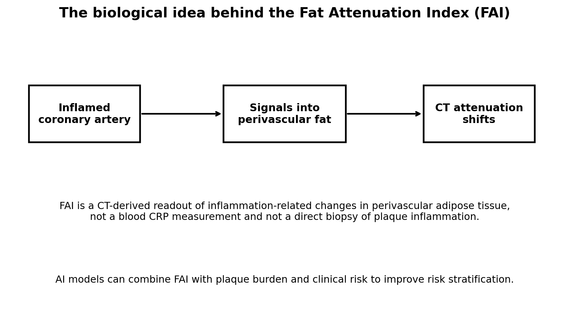
Figure 1. Coronary inflammation changes the phenotype of perivascular fat, creating a CT signal that can be quantified by FAI-type methods.
1. Why Perivascular Fat Contains Information About the Artery
Perivascular adipose tissue is biologically active. Inflammation within the adjacent vessel wall inhibits local fat-cell lipid accumulation and alters water/lipid content. CT attenuation becomes less negative in inflamed regions, allowing an imaging biomarker to be derived from a standard contrast CCTA.
2. CRISP-CT
The 2018 CRISP-CT study showed that perivascular FAI around the major coronary arteries was associated with cardiac mortality independent of traditional risk factors and conventional CCTA interpretation. The study created the modern clinical interest in coronary perivascular fat mapping.
3. ORFAN: The Large Real-World Test
ORFAN enrolled more than 40,000 consecutive patients across eight UK hospitals. The central clinical problem was obvious: most patients undergoing CCTA did not have obstructive CAD, yet events still occurred in this population.
In the publication, 81.1% of the cohort had no obstructive CAD. Because this group was large, nearly twice as many cardiac deaths and major adverse cardiac events occurred among people without obstruction as among those with obstructive disease in absolute terms during early follow-up.
In the long-term subcohort, FAI-based inflammation and an AI-enhanced risk score incorporating plaque and clinical features improved risk discrimination. This supports the idea that anatomy alone does not capture all residual risk.
4. FAI vs hsCRP
| Feature | hsCRP | FAI / perivascular fat |
|---|---|---|
| Sample | Blood | CCTA image |
| Signal | Systemic inflammation | Local coronary inflammatory environment |
| Anatomic specificity | No | Yes, around coronary arteries |
| Standard guideline use | Established risk enhancer | Emerging/advanced imaging |
| Technical dependence | Low-moderate | Scanner/reconstruction/software dependent |
5. What Should Change if FAI Is High?
The evidence supports stronger risk recognition, not an automatic drug algorithm. The first response should be to ensure LDL-C/ApoB, blood pressure, smoking, diabetes, exercise and weight are optimally managed.
Whether FAI should trigger colchicine, IL-6 inhibition or another anti-inflammatory therapy is not yet established as routine practice. Randomized FAI-guided management trials are the next step.
6. FAQ
Can every CCTA be analyzed for FAI?
Many contrast CCTA datasets contain the information, but validated analysis software and acquisition quality are required.
Is FAI guideline-mandated?
No. It is an advanced biomarker with growing evidence, not a universal required component of CCTA interpretation.
Is FAI better than plaque burden?
They measure different dimensions. ORFAN-type models work best when inflammation, plaque and clinical risk are integrated.
Should asymptomatic people get CCTA only to measure FAI?
There is not enough outcomes evidence to support broad screening CCTA solely for FAI.
References
1. Oikonomou EK, Marwan M, Desai MY, et al. Non-invasive detection of coronary inflammation using computed tomography and prediction of residual cardiovascular risk: CRISP-CT. Lancet. 2018;392:929-939.
2. Antoniades C, et al. Inflammatory risk and cardiovascular events in patients without obstructive coronary artery disease: the Oxford Risk Factors And Non-invasive imaging (ORFAN) study. Multicentre longitudinal cohort. 2024.
3. Oxford Risk Factors and Non-Invasive Imaging (ORFAN) study: AI-enhanced risk prediction publications, 2024.
4. Libby P. Inflammation in atherosclerosis. Major review literature.