Executive Summary
Coronary CT angiography already shows stenosis, plaque location and high-risk features. AI-based quantitative coronary plaque analysis, often called QCPA or AI-QCT, adds automated segmentation and numerical estimates of total, calcified and noncalcified plaque burden.
That capability matters because most future acute coronary syndromes do not necessarily arise from lesions that were severely obstructive on the earlier scan. Studies such as ICONIC showed that plaque burden and adverse plaque characteristics add prognostic information beyond stenosis alone.
By 2026, the field has moved beyond feasibility. The ACC published a dedicated scientific statement on quantitative plaque analysis, noting that multiple FDA-cleared commercial products exist and discussing possible clinical indications and reporting standards.
The same statement also states the limitation clearly: evidence remains insufficient that QCPA improves patient-management decisions and clinical outcomes. That is the line between useful measurement and overclaiming.
New 2026 registry data continue to show that AI-quantified total plaque volume is associated with adverse cardiovascular events independently of conventional risk factors and stenosis severity. Reproducibility work is also improving.
Association is still not a treatment algorithm. There is no universally validated plaque-volume threshold that says “add drug X,” no evidence that chasing small year-to-year plaque-volume changes improves outcomes, and no reason to repeat CCTA annually in asymptomatic people simply because software can compare scans.
The best use of AI plaque analysis today is as an adjunct to a clinically indicated CCTA, especially when the result can sharpen the understanding of overall atherosclerotic burden. It should not be sold as a crystal ball for the next heart attack.
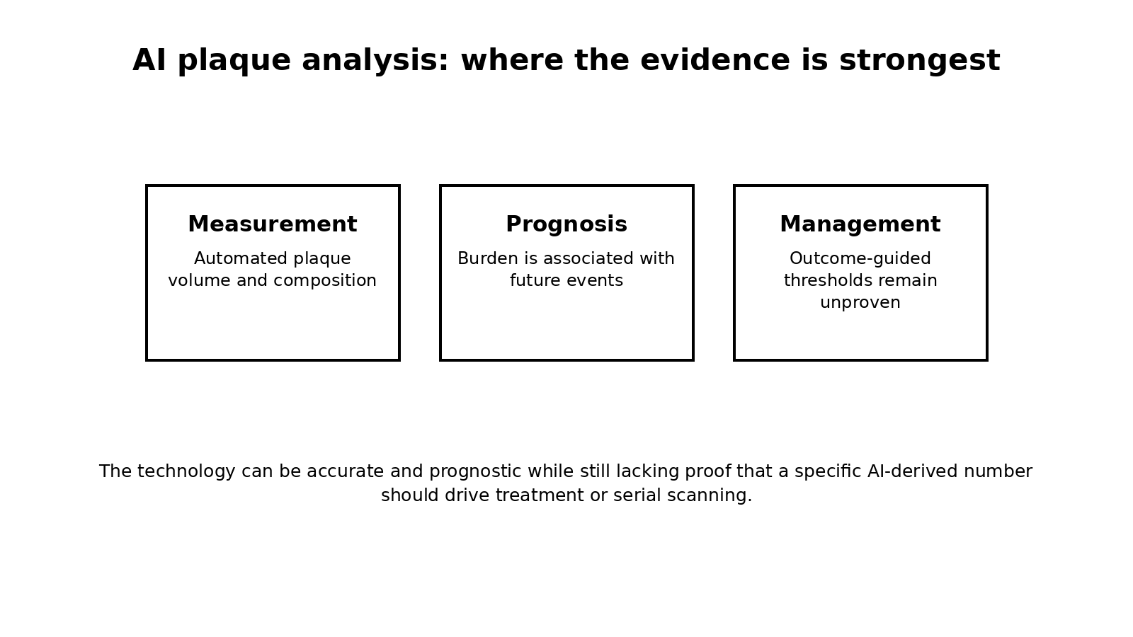
Figure 1. AI can make plaque measurement more quantitative. The evidence is strongest for measurement and prognosis, weaker for serial monitoring and treatment decisions based on the number alone.
1. What AI plaque software actually does
Modern platforms segment the coronary tree and estimate plaque volume by tissue characteristics, often separating calcified from noncalcified components and reporting percent atheroma volume or related burden metrics.
This reduces the labor of manual quantification and can improve consistency. It does not change the basic limitations of CCTA image quality, scanner technique, heart rate, artifacts or contrast timing.
2. What is already real
Plaque burden and composition contain prognostic information. ICONIC showed that many culprit precursors were nonobstructive at baseline, while adverse plaque features and burden were associated with later acute coronary syndromes.
A 2026 Mass General Brigham registry analysis reported that AI-quantified total plaque volume was associated with adverse events even after accounting for risk factors and stenosis severity. This supports prognostic validity, not yet an outcomes-guided treatment protocol.
3. What is not yet proven
A software report may give plaque volume to the cubic millimeter. Precision of output should not be confused with certainty of meaning. The ACC statement notes insufficient evidence that QCPA improves management decisions and patient outcomes.
There is also no reliable way to point to one nonobstructive plaque and guarantee that it will be the future culprit. High-risk features can identify a higher-risk patient, but lesion-level prediction remains imperfect.
4. The serial-imaging trap
Serial CCTA is attractive because it appears to turn prevention into a visible before-and-after score. The problem is that small differences can reflect acquisition, reconstruction, software version or analysis variability, not true biology.
Reproducibility research published in 2026 is important, but routine annual scanning has not been validated as a therapeutic scorecard. Radiation, iodinated contrast, incidental findings and downstream testing also matter.
5. How to use an AI plaque report intelligently
First answer the clinical question that justified CCTA. Then interpret stenosis, overall plaque burden, high-risk features and symptoms together, not as isolated software outputs.
A heavy noncalcified plaque burden can strengthen the case for aggressive risk-factor control even when CAC is low. But the treatment target remains the patient’s risk and proven causal exposures, especially LDL/ApoB, blood pressure, smoking and diabetes, not a proprietary plaque-volume threshold.
| Claim | 2026 verdict |
|---|---|
| AI can quantify plaque from CCTA | Real, commercially available and increasingly standardized |
| Plaque burden predicts risk | Supported by observational and registry evidence |
| AI identifies the exact plaque that will rupture | Not reliably proven |
| A plaque-volume cutoff dictates medication choice | Not established |
| Annual CCTA tracks treatment success | Not validated for routine asymptomatic follow-up |
| Best current role | Adjunct to clinically indicated CCTA, interpreted with the full clinical picture |
If you are offered AI plaque analysis, ask three questions: Was the CCTA clinically indicated in the first place? Which outputs are independently validated and reproducible? Most importantly, what management decision would change because of the result? A more detailed number is useful only when it improves a real decision.
6. FAQ
Is AI plaque analysis FDA cleared?
Multiple commercial quantitative plaque-analysis products have FDA clearance, according to the ACC scientific statement. Clearance supports intended technical use; it does not prove that using the output improves cardiovascular outcomes.
Can AI see soft plaque when CAC is zero?
CCTA can detect noncalcified plaque that a calcium scan cannot. AI may quantify that burden, but CCTA should still be ordered for an appropriate clinical reason rather than used reflexively after every CAC 0 result.
Should I repeat CCTA in one year to see if plaque regressed?
Usually not as a routine strategy. Serial quantitative plaque analysis is an active research area, and small changes may be within measurement variability.
Is more plaque detail always better?
No. More detail can improve risk understanding, but it can also create false precision and downstream testing if there is no validated action tied to the measurement.
References
1. Chandrashekhar Y, Blankstein R, Shaw LJ, et al. Quantitative Coronary Plaque Analysis in Clinical Practice: 2025 ACC Scientific Statement. JACC Cardiovasc Imaging. 2026;19:637-652.
2. Chang HJ, Lin FY, Lee SE, et al. Coronary Atherosclerotic Precursors of Acute Coronary Syndromes. J Am Coll Cardiol. 2018;71:2511-2522.
3. Huck D, Shiyovich A, Cardoso R, et al. AI-Based Coronary Plaque Analysis and Adverse Cardiovascular Events: The Mass General Brigham CCTA Registry. J Am Coll Cardiol. 2026;87(13 Suppl):A889-A890.
4. Kim DW, Jaltotage B, Fonte T, et al. Reproducibility of AI-Informed Coronary Computed Tomography Angiography-Derived Coronary Plaque Volume Quantification. JACC Adv. 2026. Published online July 11, 2026.