Publication metadata
| Field | Recommendation |
|---|---|
| SEO title | Inflammation and Atherosclerosis: hsCRP, Colchicine and Residual Cardiovascular Risk |
| Suggested slug | /inflammation-atherosclerosis.html |
| Primary keyphrase | inflammation and atherosclerosis |
| Meta description | A practical evidence-based guide to hsCRP, residual inflammatory risk, CANTOS, colchicine, IL-6 and CCTA-derived coronary inflammation. |
| Audience | Patients, clinicians and readers interested in preventive cardiology |
| Author | ElevatedCholesterol.com Editorial Team |
Executive summary
Atherosclerosis is neither “just cholesterol” nor “just inflammation.” The most useful modern model starts with retention of ApoB-containing lipoproteins within the arterial wall and then follows a self-amplifying inflammatory response involving endothelial activation, monocytes, macrophages, inflammasome signaling and cytokines such as interleukin-1β (IL-1β) and interleukin-6 (IL-6). The lipid substrate and the inflammatory response are biologically intertwined. [1–3]
High-sensitivity C-reactive protein (hsCRP) is the most practical blood marker for systemic inflammation in cardiovascular risk assessment, but it is not a coronary-specific test. A value around 2 mg/L or higher has been used repeatedly to define an inflammatory-risk phenotype in landmark trials, including JUPITER and CANTOS. Acute infection, trauma, autoimmune disease and other inflammatory states can raise hsCRP, so interpretation requires clinical context and, when appropriate, repeat measurement. [4–7]
The strongest proof that inflammation itself can be therapeutically relevant came from CANTOS: selectively blocking IL-1β reduced recurrent cardiovascular events without lowering LDL-C. Low-dose colchicine then produced positive outcome results in COLCOT and LoDoCo2, but the later CLEAR SYNERGY trial found no cardiovascular benefit when colchicine was started soon after myocardial infarction despite lowering CRP. The correct 2026 conclusion is therefore nuanced: inflammation is a validated treatment target, but benefit depends on the drug, population, timing and biology being targeted. [7–11]
Guidelines reflect that nuance. The 2023 US chronic coronary disease guideline states that adding low-dose colchicine may be considered for secondary prevention, while the 2024 ESC chronic coronary syndrome guideline gives a stronger recommendation for selected patients with atherosclerotic coronary disease. Colchicine is not a substitute for aggressive LDL/ApoB lowering, blood-pressure control, smoking cessation, exercise, weight management or antithrombotic therapy when indicated. [12,13]
Coronary CT is also beginning to move inflammation from a systemic biomarker to a local imaging phenotype. Perivascular fat attenuation index (FAI) uses CCTA data to detect changes in fat surrounding an inflamed coronary artery. CRISP-CT and the 40,091-patient ORFAN study showed that FAI-derived inflammatory risk can add prognostic information beyond stenosis and conventional risk factors, particularly in people without obstructive CAD. This technology is promising, but it should not yet be treated as a universal screening test or a replacement for guideline-based risk management. [14–16]
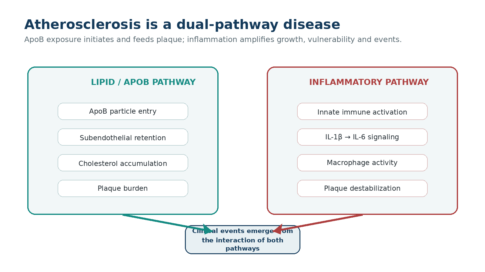
Figure 1. Atherosclerosis is best understood as an interaction between ApoB exposure and arterial-wall inflammation. Illustration created for ElevatedCholesterol.com.
At a glance
| Question | Evidence-based answer |
|---|---|
| Is inflammation causal in atherosclerosis? | Yes. Mechanistic, genetic and randomized trial evidence supports a causal role, but inflammation operates within a disease initiated and sustained by atherogenic lipoproteins. |
| Is hsCRP a “plaque inflammation test”? | No. It is a systemic downstream biomarker. It can help identify residual inflammatory risk, but it does not localize inflammation to a coronary plaque. |
| Does lowering CRP automatically prevent events? | No. CRP is primarily a marker. Different anti-inflammatory interventions have produced different outcome results. |
| Does colchicine work? | It reduced events in COLCOT and LoDoCo2, but not in CLEAR SYNERGY. Current guidelines support selective use in established coronary disease, not indiscriminate use. |
| Can CCTA image inflammation? | Emerging techniques such as perivascular FAI can estimate coronary inflammatory activity from routine CCTA data and add prognostic information, but availability and clinical integration vary. |
1. Why "cholesterol versus inflammation" is the wrong debate
For decades, public discussion often framed atherosclerosis as a cholesterol-storage problem. More recently, social media sometimes swings too far in the opposite direction and claims that plaque is “really an inflammatory disease” and cholesterol is secondary. Both simplifications are misleading.
The arterial wall does not become atherosclerotic because CRP happens to be high. The process begins when ApoB-containing particles — LDL, remnant particles and Lp(a) among them — cross the endothelium and become retained in the subendothelial space. Their lipids and associated molecular signals trigger endothelial activation and recruit monocytes. Macrophages ingest modified lipoproteins, become foam cells and help build the lipid-rich necrotic core. [1–3]
Inflammation then changes the trajectory of disease. Cytokine signaling increases immune-cell recruitment, matrix-degrading enzymes can weaken the fibrous cap, and an active inflammatory microenvironment can make plaque more prone to rupture. Inflammation also participates in healing and calcification. In other words, the same immune system that responds to arterial injury can both protect and destabilize depending on context.
This is why modern prevention increasingly thinks in terms of two residual risks. Residual lipid risk remains when ApoB, LDL-C, remnant cholesterol or Lp(a) exposure remains excessive despite treatment. Residual inflammatory risk remains when inflammatory activity stays elevated despite acceptable lipid levels and otherwise good preventive therapy. The risks overlap rather than compete.
Do not use a low hsCRP to excuse a high ApoB, or a low LDL-C to assume inflammation is irrelevant. The strongest prevention strategy controls the atherogenic substrate first and then considers whether meaningful residual inflammatory risk remains.
2. How arterial inflammation actually works
The inflammatory biology of atherosclerosis is a network rather than a single pathway. A useful simplified sequence is: ApoB retention → cholesterol crystal and danger-signal formation → inflammasome activation → IL-1β signaling → IL-6 signaling → hepatic production of CRP and other acute-phase proteins. [1,2,7]
The NLRP3 inflammasome is one of the best-studied sensors in this pathway. Within macrophages, it can respond to cholesterol crystals and other danger signals, leading to activation of IL-1β. IL-1β then promotes downstream IL-6, which in turn drives hepatic CRP production. This hierarchy explains why CRP is useful clinically but is not itself the central therapeutic target in most contemporary strategies.
Inflammation also influences the physical structure of plaque. Activated macrophages release proteases and inflammatory mediators that can thin the fibrous cap. At the same time, healing responses can increase collagen and calcium. Consequently, an artery can simultaneously contain old, densely calcified, relatively stable plaque and younger lipid-rich, inflamed plaque. That mixed biology is one reason a simple lumen measurement or calcium score cannot tell the whole story.
Lp(a) adds another layer because it carries oxidized phospholipids, which have pro-inflammatory properties and may contribute to both atherosclerosis and aortic valve disease. This helps explain why elevated Lp(a) can confer risk even when LDL-C is low. [17]
The IL-1β → IL-6 → CRP axis
| Level | What it represents | Clinical relevance |
|---|---|---|
| IL-1β | Upstream innate immune cytokine activated in part through inflammasome signaling. | CANTOS proved that directly targeting this pathway can reduce recurrent events without changing LDL-C. |
| IL-6 | Downstream cytokine with broad hepatic and vascular effects. | A major current therapeutic target; phase 3 outcomes programs are testing whether IL-6 inhibition reduces events. |
| hsCRP | Downstream hepatic acute-phase protein. | Accessible risk biomarker; useful for identifying a systemic inflammatory phenotype, but not artery-specific. |
3. hsCRP: useful biomarker, commonly misunderstood
High-sensitivity CRP is the same protein measured by standard CRP assays, but the high-sensitivity method accurately quantifies the low concentrations relevant to chronic cardiovascular risk. It is inexpensive, widely available and has a large evidence base.
In cardiovascular studies, hsCRP around 2 mg/L or higher has often been used to identify residual inflammatory risk. JUPITER enrolled apparently healthy people with LDL-C below 130 mg/dL but hsCRP of at least 2 mg/L. CANTOS required previous myocardial infarction plus hsCRP of at least 2 mg/L. PROVE IT–TIMI 22 analyses also showed that, after acute coronary syndrome, patients who achieved CRP below 2 mg/L had fewer recurrent events across achieved LDL-C strata. [4,6,7]
But hsCRP is nonspecific. Obesity, smoking, periodontal disease, acute infection, inflammatory arthritis, trauma and many other conditions can raise it. A single abnormal value should therefore not be interpreted in isolation. If hsCRP is markedly elevated — especially above about 10 mg/L — clinicians commonly look for an acute or non-cardiovascular inflammatory cause and repeat the measurement after the transient process has resolved.
The 2026 ACC/AHA dyslipidemia guideline supports selective use of hsCRP to personalize risk assessment when the decision remains uncertain rather than universal testing for everyone. [18]
Myth: “hsCRP tells me whether my coronary plaque is inflamed.” Fact: hsCRP is systemic. It can identify an inflammatory-risk phenotype, but it cannot tell which artery or plaque is inflamed. Local coronary inflammation imaging is a different technology.
4. Residual inflammatory risk: what remains after LDL is treated
The concept of residual inflammatory risk became clinically important because cardiovascular events continued to occur even in patients receiving intensive lipid-lowering therapy. Some of that risk is simply residual lipid exposure — LDL-C or ApoB not low enough, Lp(a), remnants, or years of accumulated lifetime exposure. But part of the remaining risk tracks with inflammatory biomarkers.
In PROVE IT–TIMI 22, achieved LDL-C and achieved CRP each carried prognostic information. Patients with both LDL-C below 70 mg/dL and CRP below 2 mg/L did better than patients who reached only one of those targets. In the parallel REVERSAL imaging analysis, changes in CRP and LDL-C were independently associated with coronary plaque progression. [4,5]
These findings do not mean clinicians should “treat to a CRP target” in the same mechanical way they treat to an LDL-C goal. They established a biological idea: even after lipid lowering, persistent inflammatory activity can mark people whose risk remains higher.
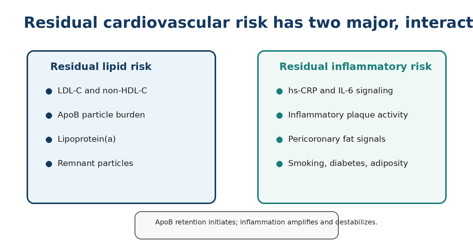
Figure 2. Residual lipid risk and residual inflammatory risk are overlapping, potentially modifiable dimensions of risk. Original ElevatedCholesterol.com figure.
5. The landmark trials: from association to causality
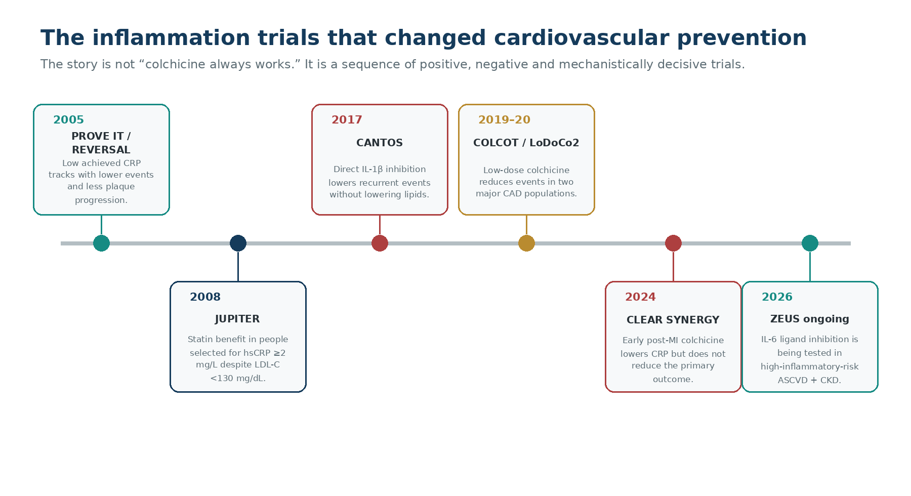
Figure 3. Landmark studies established the inflammatory hypothesis, demonstrated event reduction with selected anti-inflammatory strategies, and also showed that benefit is not universal. Original ElevatedCholesterol.com figure.
PROVE IT–TIMI 22 and REVERSAL: the “dual target” clue
The 2005 analyses from PROVE IT–TIMI 22 and REVERSAL were not trials of a dedicated anti-inflammatory drug. Their importance was conceptual. Intensive statin therapy lowered both LDL-C and CRP. In PROVE IT, patients with achieved CRP below 2 mg/L had fewer recurrent events regardless of achieved LDL-C, and those with both low LDL-C and very low CRP had the best outcomes. In REVERSAL, reductions in LDL-C and CRP were independently related to slower plaque progression. [4,5]
JUPITER: inflammation identifies risk even when LDL-C is not “high”
JUPITER randomized 17,802 apparently healthy adults with LDL-C below 130 mg/dL and hsCRP at least 2 mg/L to rosuvastatin 20 mg or placebo. Rosuvastatin lowered LDL-C by about 50% and hsCRP by about 37%; the primary cardiovascular endpoint was reduced by 44% (HR 0.56). JUPITER did not isolate inflammation from lipid lowering, but it demonstrated that people selected partly because of inflammation could benefit substantially from intensive statin therapy. [6]
CANTOS: the causal breakthrough
CANTOS provided the clearest randomized proof that inflammation is not merely a bystander. More than 10,000 people with previous MI and hsCRP at least 2 mg/L received canakinumab, a monoclonal antibody targeting IL-1β, or placebo. The 150-mg dose reduced the primary endpoint by about 15% without lowering LDL-C. Fatal infection was increased, and all-cause mortality was not reduced. The drug did not become routine cardiovascular therapy, but the biology changed preventive cardiology permanently: directly reducing a specific inflammatory pathway can reduce atherothrombotic events independently of lipid lowering. [7]
COLCOT: low-dose colchicine after MI
COLCOT randomized 4,745 patients within 30 days after myocardial infarction to colchicine 0.5 mg daily or placebo. Over a median 22.6 months, the primary composite occurred in 5.5% versus 7.1% (HR 0.77). The absolute difference was 1.6 percentage points. Pneumonia was uncommon but more frequent with colchicine in the original trial. [9]
LoDoCo2: chronic coronary disease
LoDoCo2 enrolled approximately 5,500 patients with chronic coronary disease. Over a median 28.6 months, the primary endpoint occurred in 6.8% with colchicine and 9.6% with placebo (HR 0.69), a 31% relative reduction and a 2.8-percentage-point absolute reduction. A numerically higher rate of non-cardiovascular death in the colchicine group did not reach conventional statistical significance, and the signal remains part of the safety discussion. [10]
CLEAR SYNERGY: an important negative trial
Then came a result that prevents an overly simple narrative. CLEAR SYNERGY randomized 7,062 patients with acute MI to colchicine or placebo. Over a median three years, the primary endpoint occurred in 9.1% versus 9.3% (HR 0.99; P=0.93). Colchicine lowered CRP at three months but did not lower cardiovascular events; diarrhea was more common. [11]
CLEAR SYNERGY shows why “lower inflammation = fewer events” is not a universal rule. Biomarker reduction does not guarantee clinical benefit. Timing, patient selection, background therapy, adherence, target pathway and endpoint composition all matter.
| Trial | Population | Intervention | Primary result | What it taught us |
|---|---|---|---|---|
| JUPITER (2008) | Primary prevention; LDL-C <130 mg/dL; hsCRP ≥2 mg/L | Rosuvastatin 20 mg | HR 0.56 | Inflammatory phenotype can identify people who benefit from aggressive statin therapy, but lipid and inflammation effects were intertwined. |
| CANTOS (2017) | Prior MI; hsCRP ≥2 mg/L | Canakinumab | 150 mg HR 0.85 | Direct anti-inflammatory event reduction independent of LDL lowering. |
| COLCOT (2019) | Recent MI | Colchicine 0.5 mg/d | HR 0.77 | Positive secondary-prevention signal after MI. |
| LoDoCo2 (2020) | Chronic coronary disease | Colchicine 0.5 mg/d | HR 0.69 | Strong positive result in stable CAD. |
| CLEAR SYNERGY (2024) | Acute MI | Colchicine 0.5 mg/d | HR 0.99 | Colchicine is not uniformly effective across coronary populations and treatment settings. |
6. Colchicine in 2026: where it fits --- and where it does not
Colchicine is now a legitimate cardiovascular drug, but not a universal “anti-inflammatory supplement.” Its evidence base is strongest in secondary prevention among people with established atherosclerotic coronary disease. In the United States, a 0.5-mg formulation is approved to reduce the risk of myocardial infarction, stroke, coronary revascularization and cardiovascular death in adults with established atherosclerotic disease or multiple cardiovascular risk factors. [19]
The 2023 AHA/ACC chronic coronary disease guideline states that adding colchicine for secondary prevention may be considered (Class 2b, Level B-R). The 2024 ESC chronic coronary syndrome guideline moved further, supporting low-dose colchicine more strongly in patients with atherosclerotic CAD. The difference reflects both evidence interpretation and guideline philosophy. [12,13]
CLEAR SYNERGY complicates the story but does not erase LoDoCo2 or COLCOT. It instead argues against assuming a class-wide or timing-independent effect. A patient with stable chronic coronary disease is not biologically identical to a patient treated immediately after acute MI, and trial populations differed in background therapy and follow-up.
Safety and drug interactions matter
Colchicine has a narrow therapeutic window compared with ordinary preventive medications. It is metabolized through CYP3A4 and transported by P-glycoprotein. Strong inhibitors of these pathways can cause dangerous increases in colchicine exposure. Severe renal or hepatic dysfunction also changes safety substantially. The US prescribing information specifically contraindicates important combinations and warns about neuromuscular toxicity and rhabdomyolysis; gastrointestinal symptoms are among the common early signs of excess exposure. [19]
| Question | Practical 2026 answer |
|---|---|
| Should everyone with a high hsCRP take colchicine? | No. There is no validated “hsCRP ≥2 → prescribe colchicine” algorithm. |
| Should colchicine replace LDL/ApoB lowering? | No. It addresses a different pathway and was tested on top of standard preventive therapy. |
| Is it a primary-prevention wellness drug? | No. Routine use in low-risk people without established disease is not supported by current cardiovascular outcomes evidence. |
| Can it interact with common drugs? | Yes. CYP3A4/P-gp interactions, renal/hepatic function and myotoxicity risk require medication review. |
7. IL-6: the next major therapeutic test
If CANTOS established IL-1β as a causal target, IL-6 is the next major node in the pathway. Genetic, epidemiologic and trial data make IL-6 signaling an attractive target because it lies downstream of IL-1β and upstream of hepatic CRP production.
Ziltivekimab is a monoclonal antibody that neutralizes the IL-6 ligand. Phase 2 work showed very large reductions in hsCRP and other inflammatory biomarkers in people with chronic kidney disease and elevated inflammatory risk. The phase 3 ZEUS cardiovascular outcomes trial is testing whether those biomarker changes translate into fewer major events in patients with established ASCVD, CKD and hsCRP at least 2 mg/L. As of 1 August 2026, ZEUS remains an outcomes trial in progress; it should not be described as proven cardiovascular therapy. [20]
Other IL-6–pathway programs are also being studied in acute MI, heart failure and kidney disease. The crucial lesson from CLEAR SYNERGY is that phase 3 outcome data matter more than biomarker enthusiasm.
IL-6 inhibition is one of the most important emerging strategies in residual inflammatory risk, but cardiovascular event reduction with ziltivekimab has not yet been established as of this publication date.
8. Can we image coronary inflammation? FAI, CCTA and AI risk models
Blood biomarkers tell us about systemic inflammation. Coronary CT may be able to provide something different: a local signature of inflammatory activity surrounding individual coronary arteries.
Inflamed coronary arteries release signaling molecules that alter the biology of adjacent perivascular adipose tissue. This changes adipocyte size and lipid content, which changes CT attenuation. The perivascular fat attenuation index (FAI) was developed to quantify this gradient on CCTA. [14]
In CRISP-CT, FAI added prognostic information for cardiac mortality beyond traditional risk factors, coronary anatomy and high-risk plaque features. The 2024 ORFAN study then evaluated 40,091 consecutive patients undergoing clinically indicated CCTA across eight UK hospitals. Most had no obstructive CAD, yet this group accounted for the majority of events. FAI-derived inflammatory risk predicted cardiac mortality and major adverse events independently of conventional risk factors and CAD extent. An AI-Risk model integrating FAI, plaque metrics and clinical factors also stratified long-term risk. [14–16]
This is scientifically important because stenosis alone is an incomplete description of risk. Many future events arise in people without severe obstruction. But FAI is not a reason to perform CCTA in every asymptomatic person, and commercially available analysis is not uniformly accessible or incorporated into all guidelines. It should be viewed as an emerging layer of precision risk assessment rather than a universal replacement for hsCRP, CAC, conventional CCTA interpretation or clinical judgment.
| Tool | What it measures | Strength | Major limitation |
|---|---|---|---|
| hsCRP | Systemic downstream inflammatory signal | Cheap, validated, widely available | Nonspecific; cannot localize coronary inflammation |
| CCTA plaque analysis | Anatomic plaque burden and composition | Direct coronary anatomy | Plaque phenotype is not the same as active inflammation |
| Perivascular FAI | CT signature in fat surrounding coronary arteries | Local coronary inflammatory phenotype; prognostic data | Specialized analysis; evolving clinical integration and access |
| PET inflammation imaging | Tracer uptake related to metabolic/inflammatory activity | Research-grade biological imaging | Cost, radiation, resolution and limited routine coronary use |
9. Where Lp(a) fits into the inflammatory story
Lp(a) is often discussed as a lipid risk factor, but that description is incomplete. Its LDL-like particle carries ApoB and cholesterol, while the apo(a) component preferentially binds oxidized phospholipids. These oxidized phospholipids can activate inflammatory pathways in vascular and valvular tissue. [17]
This dual biology is one reason high Lp(a) can remain clinically important even when conventional LDL-C is aggressively controlled. Current management therefore focuses on reducing all modifiable risk — especially LDL-C/ApoB exposure, blood pressure, smoking and metabolic risk — while dedicated Lp(a)-lowering outcome trials continue to define how much event reduction can be achieved by targeting Lp(a) itself.
An elevated hsCRP does not prove that Lp(a) is causing an individual patient’s inflammation, and a low hsCRP does not neutralize the genetically mediated risk associated with high Lp(a). They are different measurements of different biological dimensions.
10. Lifestyle: anti-inflammatory without the marketing language
The most defensible “anti-inflammatory protocol” is not a supplement stack. It is aggressive control of the exposures known to drive both vascular risk and systemic inflammation.
Regular physical activity, smoking cessation, weight reduction when excess adiposity is present, high-quality dietary patterns, adequate sleep and treatment of diabetes and periodontal or inflammatory disease can all lower inflammatory burden while improving other cardiovascular pathways. The Mediterranean dietary pattern has randomized outcomes evidence for cardiovascular prevention; exercise improves endothelial function and cardiorespiratory fitness; smoking cessation removes a potent vascular inflammatory stimulus.
The important editorial distinction is between lowering a biomarker and proving fewer cardiovascular events. Curcumin, nattokinase, serrapeptase, “glycocalyx” supplements and many other products are frequently marketed as anti-inflammatory or plaque-regressing therapies, but they do not have an evidence base remotely comparable with statins, blood-pressure treatment, smoking cessation, exercise, selected GLP-1 therapies, colchicine in appropriate coronary disease, or the established antithrombotic and lipid-lowering strategies used in guidelines.
Prioritize the interventions with outcome data: do not smoke; move regularly; improve fitness; manage weight and glycemia; use a Mediterranean-style dietary pattern; treat LDL/ApoB aggressively according to risk; control blood pressure; and discuss any drug aimed at residual inflammatory risk with a clinician who can review kidney/liver function and interactions.
11. A practical way to use hsCRP
Measure hsCRP when the result could change how you think about risk — particularly when risk appears discordant or residual inflammatory risk is clinically relevant.
Interpret it only when the patient is clinically stable. Fever, infection, recent surgery, acute injury and active inflammatory disease can dominate the result.
If the value is unexpectedly high, repeat it after transient inflammatory conditions have resolved rather than labeling the patient “vascularly inflamed” from one measurement.
Use hsCRP alongside — not instead of — LDL-C, ApoB, Lp(a), blood pressure, diabetes status, smoking, family history and, when appropriate, CAC/CCTA.
In established coronary disease, persistent inflammatory risk can prompt a clinician to review whether all foundational therapies are optimized and whether guideline-supported low-dose colchicine is appropriate.
Do not chase hsCRP with unproven supplements simply to normalize a laboratory number.
A simple decision framework
| Clinical situation | Reasonable interpretation |
|---|---|
| Low ApoB / LDL-C + low hsCRP | Both major residual pathways appear relatively controlled; continue overall risk management. |
| High ApoB / LDL-C + low hsCRP | Residual lipid risk remains the priority. A low hsCRP does not neutralize ApoB exposure. |
| Low ApoB / LDL-C + hsCRP persistently ≥2 mg/L | Consider residual inflammatory risk, rule out nonvascular causes, and in established CAD review guideline-supported anti-inflammatory options. |
| High ApoB / LDL-C + high hsCRP | Both pathways remain active. Treat foundational risk factors first and comprehensively. |
12. Myth vs fact
| Myth | Fact |
|---|---|
| “Plaque is not a cholesterol problem — it is an inflammation problem.” | False dichotomy. ApoB retention is a causal driver; inflammation is a crucial mediator of progression and complications. |
| “If my hsCRP is low, my arteries are safe.” | False. Low hsCRP does not rule out significant plaque, high ApoB exposure, elevated Lp(a), diabetes, hypertension or genetic risk. |
| “If hsCRP is high, colchicine is the answer.” | False. High hsCRP is nonspecific, and colchicine use depends on clinical disease, safety and guideline context. |
| “CANTOS proved CRP itself causes heart attacks.” | No. CANTOS targeted IL-1β, an upstream inflammatory pathway. CRP was a downstream biomarker. |
| “Colchicine cut events by 31%, so it works for everyone.” | No. The 31% relative reduction comes from LoDoCo2 in chronic coronary disease. CLEAR SYNERGY was neutral after acute MI. |
| “AI inflammation imaging can replace standard CCTA interpretation.” | No. FAI and AI-derived models can add information, but anatomy, plaque burden, symptoms and established risk factors still matter. |
13. Frequently asked questions
What is a "good" hsCRP?
There is no single number that diagnoses coronary safety. In preventive cardiology, \<1 mg/L is often considered a low inflammatory range, 1--3 mg/L intermediate and \>3 mg/L higher, while ≥2 mg/L has been widely used as a risk-enhancing / trial-enrollment threshold. Context and repeat testing matter.
Should hsCRP be measured every year?
Not routinely for everyone. It is most useful when it can refine risk, investigate discordance, or characterize residual risk in selected patients.
Can statins lower inflammation?
Yes. Statins typically lower hsCRP as well as LDL-C. Their proven cardiovascular benefit should not be reduced to a single mechanism; lowering atherogenic lipoproteins remains foundational.
Does colchicine lower LDL-C?
No meaningful LDL-C reduction is expected. It targets inflammatory pathways and is used, when appropriate, on top of lipid-lowering therapy.
Is colchicine safe with a statin?
Often, but not automatically. Drug interactions, kidney/liver function and neuromuscular toxicity risk must be reviewed, especially with CYP3A4 or P-gp inhibitors.
Why did CLEAR SYNERGY fail if LoDoCo2 worked?
The trials studied different populations and treatment contexts. The neutral result is a reminder that timing and disease state matter and that a biomarker response does not guarantee fewer events.
Can I lower inflammation with diet alone?
A high-quality dietary pattern can improve inflammatory and metabolic health, but diet should not be used as a substitute for indicated lipid-lowering, blood-pressure or antithrombotic therapy.
Is FAI available on every CCTA?
The raw CCTA contains the image data, but validated FAI analysis requires specialized software and workflows and is not universally available.
Does high Lp(a) mean hsCRP will be high?
Not necessarily. Lp(a) carries pro-inflammatory oxidized phospholipids, but circulating hsCRP and Lp(a) measure different aspects of biology and can be discordant.
What is the next big anti-inflammatory trial?
The ZEUS outcomes program testing IL-6 ligand inhibition with ziltivekimab is one of the major ongoing phase 3 tests as of August 2026.
14. Key take-home messages
Atherosclerosis requires both an atherogenic lipoprotein substrate and an inflammatory arterial response; “cholesterol versus inflammation” is the wrong framing.
hsCRP is a useful systemic biomarker, not a coronary-plaque imaging test.
CANTOS proved that selectively reducing inflammation can reduce cardiovascular events independently of LDL lowering.
Colchicine has positive evidence in chronic CAD and selected post-MI populations, but CLEAR SYNERGY shows that benefit is not universal.
Low-dose colchicine is guideline-supported for selected patients with established coronary disease, with stronger endorsement in the 2024 ESC CCS guideline than in the 2023 US CCD guideline.
Drug interactions and renal/hepatic function are central to colchicine safety.
IL-6 inhibition is a major emerging strategy; outcomes evidence is still awaited as of 1 August 2026.
Perivascular FAI and AI-enhanced CCTA can quantify a local coronary inflammatory phenotype and may improve risk prediction, but they remain an adjunct rather than a universal screening strategy.
Lipid lowering remains foundational. Treating inflammation does not make LDL-C, ApoB or Lp(a) irrelevant.
The practical goal is not to normalize one biomarker. It is to reduce the total probability of plaque progression, rupture and clinical events.
Bibliography verification: journal/PubMed/official-source metadata checked 7 August 2026; independent clinical review remains pending.
References
1. Libby P. Inflammation in atherosclerosis. Nature. 2002;420:868–874.
2. Hansson GK. Inflammation, atherosclerosis, and coronary artery disease. N Engl J Med. 2005;352:1685–1695.
3. Borén J, Chapman MJ, Krauss RM, et al. Low-density lipoproteins cause atherosclerotic cardiovascular disease: pathophysiological, genetic, and therapeutic insights. Eur Heart J. 2020;41:2313–2330.
4. Ridker PM, Cannon CP, Morrow D, et al. C-reactive protein levels and outcomes after statin therapy. N Engl J Med. 2005;352:20–28. doi:10.1056/NEJMoa042378.
5. Nissen SE, Tuzcu EM, Schoenhagen P, et al. Statin therapy, LDL cholesterol, C-reactive protein, and coronary artery disease. N Engl J Med. 2005;352:29–38. doi:10.1056/NEJMoa042000.
6. Ridker PM, Danielson E, Fonseca FAH, et al. Rosuvastatin to prevent vascular events in men and women with elevated C-reactive protein. N Engl J Med. 2008;359:2195–2207. doi:10.1056/NEJMoa0807646.
7. Ridker PM, Everett BM, Thuren T, et al. Antiinflammatory therapy with canakinumab for atherosclerotic disease. N Engl J Med. 2017;377:1119–1131. doi:10.1056/NEJMoa1707914.
8. Ridker PM, MacFadyen JG, Everett BM, et al. Relationship of C-reactive protein reduction to cardiovascular event reduction following treatment with canakinumab: a secondary analysis from CANTOS. Lancet. 2018;391:319–328.
9. Tardif J-C, Kouz S, Waters DD, et al. Efficacy and safety of low-dose colchicine after myocardial infarction. N Engl J Med. 2019;381:2497–2505. doi:10.1056/NEJMoa1912388.
10. Nidorf SM, Fiolet ATL, Mosterd A, et al. Colchicine in patients with chronic coronary disease. N Engl J Med. 2020;383:1838–1847. doi:10.1056/NEJMoa2021372.
11. Jolly SS, d'Entremont MA, Lee SF, et al; CLEAR Investigators. Colchicine in Acute Myocardial Infarction. N Engl J Med. 2025;392(7):633-642. doi:10.1056/NEJMoa2405922.
12. Virani SS, Newby LK, Arnold SV, et al. 2023 AHA/ACC/ACCP/ASPC/NLA/PCNA guideline for the management of patients with chronic coronary disease. Circulation. 2023. doi:10.1161/CIR.0000000000001168.
13. Vrints C, Andreotti F, Koskinas KC, et al. 2024 ESC Guidelines for the management of chronic coronary syndromes. Eur Heart J. 2024;45(36):3415-3537. doi:10.1093/eurheartj/ehae177.
14. Oikonomou EK, Marwan M, Desai MY, et al. Non-invasive detection of coronary inflammation using computed tomography and prediction of residual cardiovascular risk (CRISP-CT). Lancet. 2018;392:929–939. doi:10.1016/S0140-6736(18)31114-0.
15. Antonopoulos AS, Sanna F, Sabharwal N, et al. Detecting human coronary inflammation by imaging perivascular fat. Sci Transl Med. 2017;9:eaal2658.
16. Chan K, Wahome E, Tsiachristas A, et al; ORFAN Consortium. Inflammatory risk and cardiovascular events in patients without obstructive coronary artery disease: the ORFAN multicentre, longitudinal cohort study. Lancet. 2024;403:2606–2618. doi:10.1016/S0140-6736(24)00596-8.
17. Kronenberg F, Mora S, Stroes ESG, et al. Lipoprotein(a) in atherosclerotic cardiovascular disease and aortic stenosis: a European Atherosclerosis Society consensus statement. Eur Heart J. 2022;43(39):3925-3946. doi:10.1093/eurheartj/ehac361.
18. Blumenthal RS, Morris PB, Gaudino M, et al. 2026 ACC/AHA/AACVPR/ABC/ACPM/ADA/AGS/APhA/ASPC/NLA/PCNA Guideline on the Management of Dyslipidemia. Circulation. 2026;153(17):e1154-e1276. doi:10.1161/CIR.0000000000001423.
19. US Food and Drug Administration. LODOCO (colchicine) 0.5 mg tablets: Prescribing Information. 2023. NDA 215727.
20. Fahed J, Zahid S, Blumenthal RS. IL-6 as a Predictor of CV Risk Assessment: Role of Canakinumab and Ziltivekimab in Preventive Cardiology. American College of Cardiology. July 22, 2026. https://www.acc.org/Latest-in-Cardiology/Articles/2026/07/21/12/01/IL-6-as-a-Predictor-of-CV-Risk-Assessment.
Medical disclaimer
This publication is intended for education and general information only. It is not medical advice, diagnosis or treatment and does not replace individualized care from a qualified clinician. Decisions about hsCRP testing, colchicine, anti-inflammatory therapy, lipid-lowering therapy or coronary imaging should account for the full clinical context, contraindications, drug interactions, kidney and liver function, bleeding/thrombotic risk where relevant, and current local guidelines.