Executive Summary
Aortic stenosis is usually diagnosed and followed with echocardiography. CT calcium scoring becomes particularly valuable when valve area, gradient and flow do not agree—for example in low-flow, low-gradient aortic stenosis.
The 2021 ESC/EACTS guideline uses sex-specific Agatston thresholds. Severe AS is “likely” above about 1,200 AU in women and 2,000 AU in men, and “highly likely” above about 1,600 AU in women and 3,000 AU in men. Values below about 800 AU in women and 1,600 AU in men make severe AS unlikely.
The test is non-contrast and usually ECG-gated. It answers a structural question—how heavily calcified is the valve—not a coronary question and not a direct flow question.
AVC is not a treatment-response scoreboard. A high value supports the diagnosis and prognosis of calcific valve disease, but treatment decisions still depend on symptoms, echocardiographic severity, ventricular function and the overall valve-heart interaction.
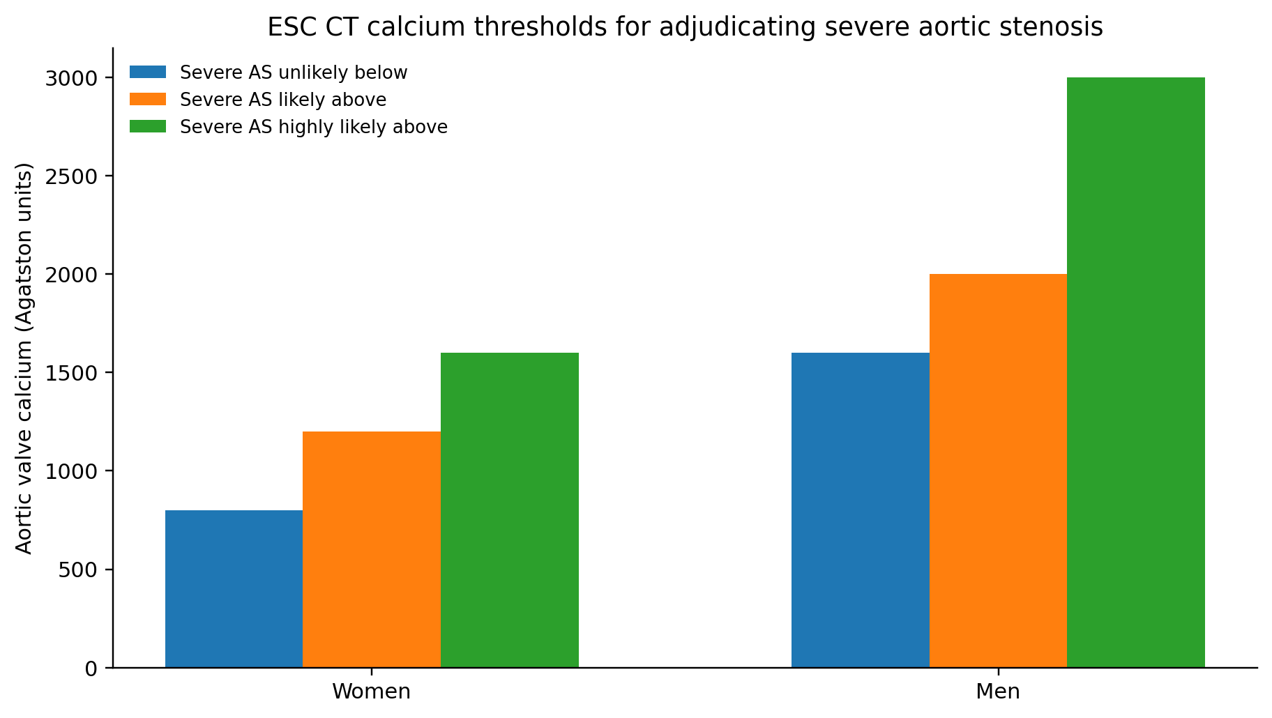
Figure 1. Sex-specific CT aortic-valve calcium thresholds used by ESC/EACTS to help adjudicate severe aortic stenosis.
1. Coronary calcium and aortic-valve calcium are different tests
Coronary CAC quantifies calcified atherosclerotic plaque in the coronary arteries. Aortic valve calcium scoring uses the same Agatston concept but applies it to the cusps of the aortic valve. One estimates coronary plaque burden; the other helps quantify calcific valve disease.
It is common for a cardiac CT report to mention both coronary calcium and aortic valve calcium, but one number cannot substitute for the other.
2. Why echocardiography remains the first-line test
Echocardiography measures peak aortic velocity, mean pressure gradient, aortic valve area, stroke volume and ventricular function. These variables determine whether the obstruction is hemodynamically important.
CT is most helpful when those variables are discordant—for example, a valve area ≤1.0 cm² but a mean gradient below 40 mmHg. In that setting, valve calcium can provide an anatomic anchor that is independent of flow.
3. Why women and men have different thresholds
Women often develop a greater fibrotic component and can reach severe hemodynamic obstruction with less total calcium than men. Using a single calcium threshold for both sexes would therefore underdiagnose severe disease in some women.
This is why guideline thresholds are sex-specific rather than simply scaled to body size.
4. How to read the thresholds
Thresholds are probabilities, not biological cliff edges. A woman with 1,190 AU is not suddenly “non-severe” because she is 10 units below 1,200, and a man with 2,010 AU is not automatically a procedural candidate. CT findings must be integrated with symptoms and Doppler data.
The “likely” and “highly likely” ranges are most useful in low-gradient cases where clinical uncertainty is real.
5. What AVC can and cannot tell you
AVC is strong for calcific burden and severity adjudication. It does not measure symptoms, left-ventricular response, coronary ischemia, bicuspid leaflet morphology in all cases, or whether an individual will benefit from intervention today.
It also does not identify the cause of every valve lesion. Rheumatic disease, endocarditis and congenital bicuspid disease can create additional structural problems beyond calcification.
6. Does Lp(a) matter?
Yes. Elevated Lp(a) is causally linked to calcific aortic valve disease and incident degenerative aortic stenosis. The association provides a biologic reason to pay attention to valve disease in patients with markedly high Lp(a), but it does not mean every such patient needs serial CT.
As of 2026, dedicated trials are testing whether large Lp(a) reduction can slow mild-to-moderate calcific AS. No Lp(a)-lowering drug has yet proved that it delays valve replacement.
| CT AVC result | Women | Men | Interpretation |
|---|---|---|---|
| Below “unlikely” threshold | <800 AU | <1,600 AU | Severe calcific AS unlikely |
| Likely severe | >1,200 AU | >2,000 AU | Supports severe AS when echo is discordant |
| Highly likely severe | >1,600 AU | >3,000 AU | Strong anatomic support for severe AS |
7. Frequently Asked Questions
Is aortic valve calcium the same as CAC?
No. Coronary CAC scores the coronary arteries; AVC scores the aortic valve.
Do I need contrast?
Standard Agatston AVC scoring is performed on a non-contrast CT.
Can a high valve calcium score diagnose severe AS by itself?
It is a powerful adjunct, especially in discordant low-gradient AS, but the diagnosis still integrates echo and clinical findings.
Can K2 lower aortic valve calcium?
A 2022 randomized MK-7 plus vitamin D trial did not slow aortic-valve calcification.
When is valve replacement needed?
Intervention depends primarily on severe AS plus symptoms, ventricular consequences, exercise testing and guideline criteria—not on calcium alone.
References
1. Vahanian A, Beyersdorf F, Praz F, et al. 2021 ESC/EACTS Guidelines for the management of valvular heart disease. Eur Heart J. 2022;43:561-632.
2. Otto CM, Nishimura RA, Bonow RO, et al. 2020 ACC/AHA Guideline for the Management of Patients With Valvular Heart Disease. Circulation. 2021;143:e72-e227.
3. Clavel MA, Messika-Zeitoun D, Pibarot P, et al. The complex nature of discordant severe calcified aortic valve disease grading. J Am Coll Cardiol.
4. Pawade T, Clavel MA, Tribouilloy C, et al. Computed tomography aortic valve calcium scoring in patients with aortic stenosis. Circ Cardiovasc Imaging. 2018.
5. Dweck MR, Boon NA, Newby DE. Calcific aortic stenosis: a disease of the valve and the myocardium. J Am Coll Cardiol. 2012;60:1854-1863.
6. Kronenberg F, Mora S, Stroes ESG, et al. Lipoprotein(a) in ASCVD and aortic stenosis: EAS consensus. Eur Heart J. 2022;43:3925-3946.
7. Diederichsen ACP, Lindholt JS, Möller S, et al. Vitamin K2 and D in patients with aortic valve calcification. Circulation. 2022;145:1387-1397.